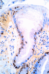
Expression of the p53 homologue p63alpha and DeltaNp63alpha in the neoplastic sequence of Barrett's oesophagus: correlation with morphology and p53 protein
- PMID: 11600462
- PMCID: PMC1728522
- DOI: 10.1136/gut.49.5.618
Expression of the p53 homologue p63alpha and DeltaNp63alpha in the neoplastic sequence of Barrett's oesophagus: correlation with morphology and p53 protein
Abstract
Background: While loss of p53 function is a key oncogenic step in human tumorigenesis, mutations of p53 are generally viewed as late events in the metaplasia-dysplasia-adenocarcinoma sequence of Barrett's oesophagus. Recent reports of a series of genes (p63, p73, and others) exhibiting close homology to p53 raise the possibility that abnormalities of these p53 family members may exert their influence earlier in the sequence.
Aim: Following recent characterisation of expression of p63 and a major isoform DeltaNp63 by generation of an antiserum that recognises p63 isoforms, but not p53, our aim was a comparative study of expression of p63 protein and p53 protein in a morphologically well defined biopsy series representative of all stages of the metaplasia-dysplasia-carcinoma sequence in Barrett's oesophagus.
Methods: A series of 60 biopsy cases representing normal oesophagus through to invasive adenocarcinoma were stained, using immunohistochemistry, with antibodies to p63 and p53. All biopsies derived from patients with endoscopic and histopathological substantiation of a diagnosis of traditional/classical Barrett's oesophagus.
Results: There was exact concordance in p53 and p63 expression in more advanced forms of neoplasia, high grade dysplasia, and invasive adenocarcinoma, while p63, but not p53, was detected in the proliferative compartment of some non-neoplastic oesophageal tissue, in both squamous mucosa and in the non-neoplastic metaplastic glandular epithelium.
Conclusions: In neoplastic Barrett's oesophagus there is upregulation of both p63 and p53 while p63 isoforms may well have an important role in epithelial biology in both non-metaplastic and metaplastic mucosa of the oesophagus. While abnormalities of p53 function represent an indisputable and critical element of neoplastic transformation, other closely linked genes and their proteins have a role in both the physiology and pathophysiology of the oesophageal mucosa.
Figures


Similar articles
-
Expression of p53-related protein p63 in the gastrointestinal tract and in esophageal metaplastic and neoplastic disorders.Hum Pathol. 2001 Nov;32(11):1157-65. doi: 10.1053/hupa.2001.28951. Hum Pathol. 2001. PMID: 11727253
-
Patterns of p53 immunoreactivity in non-neoplastic and neoplastic Barrett's mucosa of the oesophagus: in-depth evaluation in endoscopic mucosal resections.Pathology. 2019 Apr;51(3):253-260. doi: 10.1016/j.pathol.2018.12.415. Epub 2019 Feb 28. Pathology. 2019. PMID: 30826014
-
Malignant degeneration of Barrett's esophagus: the role of the Ki-67 proliferation fraction, expression of E-cadherin and p53.Dis Esophagus. 2004;17(4):322-7. doi: 10.1111/j.1442-2050.2004.00434.x. Dis Esophagus. 2004. PMID: 15569371
-
Barrett's oesophagus: from metaplasia to dysplasia and cancer.Gut. 2005 Mar;54 Suppl 1(Suppl 1):i6-12. doi: 10.1136/gut.2004.041525. Gut. 2005. PMID: 15711008 Free PMC article. Review.
-
Barrett's oesophagus--a pathologist's view.Histopathology. 2007 Jan;50(1):3-14. doi: 10.1111/j.1365-2559.2006.02569.x. Histopathology. 2007. PMID: 17204017 Review.
Cited by
-
Expression and regulation of the ΔN and TAp63 isoforms in salivary gland tumorigenesis clinical and experimental findings.Am J Pathol. 2011 Jul;179(1):391-9. doi: 10.1016/j.ajpath.2011.03.037. Epub 2011 May 7. Am J Pathol. 2011. PMID: 21703418 Free PMC article.
-
The cyclical hit model: how paligenosis might establish the mutational landscape in Barrett's esophagus and esophageal adenocarcinoma.Curr Opin Gastroenterol. 2019 Jul;35(4):363-370. doi: 10.1097/MOG.0000000000000540. Curr Opin Gastroenterol. 2019. PMID: 31021922 Free PMC article. Review.
-
Review: Experimental models for Barrett's esophagus and esophageal adenocarcinoma.Am J Physiol Gastrointest Liver Physiol. 2012 Jun 1;302(11):G1231-43. doi: 10.1152/ajpgi.00509.2011. Epub 2012 Mar 15. Am J Physiol Gastrointest Liver Physiol. 2012. PMID: 22421618 Free PMC article. Review.
-
Gain-of-function hot spot mutant p53R248Q regulation of integrin/FAK/ERK signaling in esophageal squamous cell carcinoma.Transl Oncol. 2021 Jan;14(1):100982. doi: 10.1016/j.tranon.2020.100982. Epub 2020 Dec 11. Transl Oncol. 2021. PMID: 33395748 Free PMC article.
-
Are Gastric and Esophageal Metaplasia Relatives? The Case for Barrett's Stemming from SPEM.Dig Dis Sci. 2018 Aug;63(8):2028-2041. doi: 10.1007/s10620-018-5150-0. Dig Dis Sci. 2018. PMID: 29948563 Free PMC article. Review.
References
Publication types
MeSH terms
Substances
LinkOut - more resources
Full Text Sources
Research Materials
Miscellaneous
