. 2001 Nov;49(5):692-8.
doi: 10.1136/gut.49.5.692.

Inhibitory motor innervation of the gall bladder musculature by intrinsic neurones containing nitric oxide in the Australian brush-tailed possum (Trichosurus vulpecula)

Affiliations

Inhibitory motor innervation of the gall bladder musculature by intrinsic neurones containing nitric oxide in the Australian brush-tailed possum (Trichosurus vulpecula)

A C Meedeniya et al. Gut. 2001 Nov.

Abstract

Background: Gall bladder functions are modulated by neurones intrinsic to the organ. Data are available on the neurochemical composition of intrinsic and extrinsic nerves innervating the gall bladder but are lacking on specific functional classes of gall bladder neurones.

Aims: To characterise the intrinsic motor neurones of the gall bladder and identify their roles using pharmacological techniques.

Methods: Retrograde tracing from the possum gall bladder muscle in vitro allowed identification of intrinsic motor neurones. Subsequently, their content of choline acetyltransferase and nitric oxide synthase, markers of acetylcholine and nitric oxide containing neurones, was established using immunohistochemical techniques. Organ bath pharmacology was used to evaluate neurotransmission by acetylcholine and nitric oxide in gall bladder muscle strips.

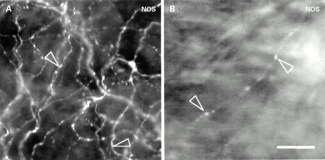
Results: Innervation of the gall bladder musculature by neurones of both the muscular and serosal plexuses was demonstrated. A large proportion (62%) of these motor neurones were immunoreactive for nitric oxide synthase. All gall bladder neurones showed immunoreactivity for choline acetyltransferase. Organ bath pharmacology confirmed the neuroanatomical data, showing acetylcholine and nitric oxide mediating neurotransmission to the gall bladder musculature.

Conclusions: Neurones containing acetylcholine and nitric oxide, located within the muscular and serosal plexuses, provide excitatory and inhibitory motor innervation of the gall bladder, respectively. The large inhibitory innervation suggests active relaxation of the gall bladder during filling, mediated by intrinsic nerves.


Figures

Figure 1
Figure 1
Photomicrographs of a ganglion of the muscular plexus in the possum gall bladder. All three neurones show choline acetyltransferase (ChAT) immunoreactivity (A). A neurone from the same ganglion shows nitric oxide synthase (NOS) immunoreactivity (arrow) while the others are immunonegative for NOS (arrowheads) (B). Bar, 20 µm.
Figure 2
Figure 2
Photomicrograph of nitric oxide synthase (NOS) immunoreactive nerve fibres within the possum gall bladder. NOS immunoreactive nerve fibres (arrowheads) were present within the non-ganglionated subepithelial plexus (A) and the muscle (B) of the gall bladder. Bar, 20 µm.
Figure 3
Figure 3
Retrogradely labelled neurones were found within the muscular (A) and serosal plexuses (C) on 1'-didodecyl-3,3,3'3'-tetra-methyl-indocarbocyanine perchlorate (DiI) application to the gall bladder muscle. Some DiI labelled cells showed immunoreactivity for nitric oxide synthase (NOS) (arrows) while others were immunonegative (arrowhead) (B, D). Bar, 20 µm.
Figure 4
Figure 4
(A) Scatterplots of neurones retrogradely labelled on 1'-didodecyl-3,3,3'3'-tetra-methyl-indocarbocyanine perchlorate (DiI) application to the gall bladder muscle in a representative preparation. The DiI labelled neurones were primarily located close to the application site (all within 10 mm). (B) Cumulative data of all DiI traced neurones from five preparations showed that nitric oxide synthase (NOS) immunoreactive neurones within both the muscular and serosal plexus were polarised in their distribution along the longitudinal axis of the gall bladder, projecting predominantly from the neck towards the apex, but NOS immunonegative cells did not show a polarised distribution.
Figure 5
Figure 5
Representative traces and cumulative data of gall bladder muscle strip pharmacology. Electrical field stimulation EFS (arrows) produced consistent gall bladder contraction under control conditions (A). Under non-adrenergic non-cholinergic (NANC) conditions, EFS produced relaxation (B). This response was abolished by N omega-nitro-L-arginine methyl ester (L-NAME) (C). Cumulative data (D) are expressed as mean (SEM). All responses differed significantly (*p<0.05) from each other. The broken lines and double headed arrows illustrate the parameters used for quantifying the EFS induced contractions (A) and relaxations (B).

References

    1. Aust N Z J Surg. 1986 Feb;56(2):147-51 - PubMed
    1. J Surg Res. 2000 Feb;88(2):186-92 - PubMed
    1. Neuroscience. 1991;42(3):863-78 - PubMed
    1. HPB Surg. 1993;7(2):125-39; discussion 139-40 - PubMed
    1. Naunyn Schmiedebergs Arch Pharmacol. 1993 Nov;348(5):526-35 - PubMed

Publication types

MeSH terms