

Loss of interstitial cells of Cajal and development of electrical dysfunction in murine small bowel obstruction
- PMID: 11600689
- PMCID: PMC2278884
- DOI: 10.1111/j.1469-7793.2001.0555c.xd
Loss of interstitial cells of Cajal and development of electrical dysfunction in murine small bowel obstruction
Abstract
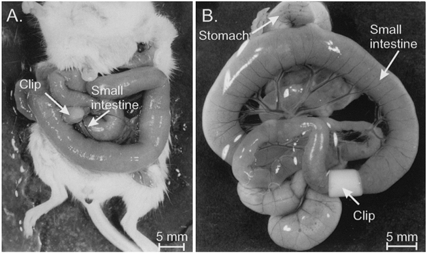
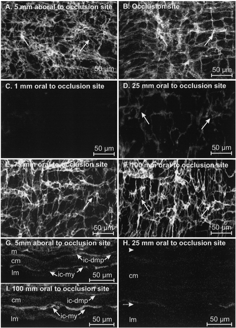
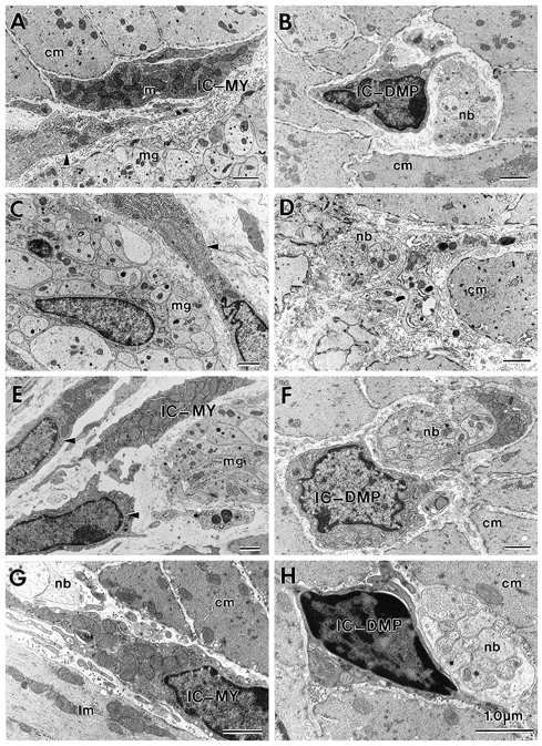
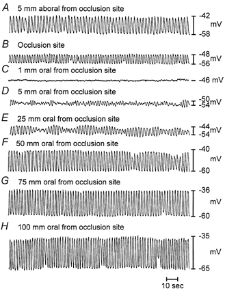
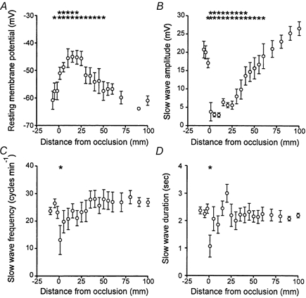
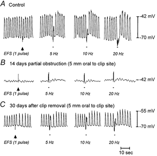
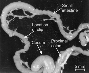
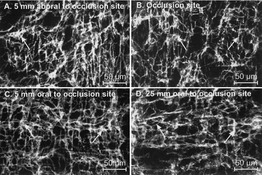
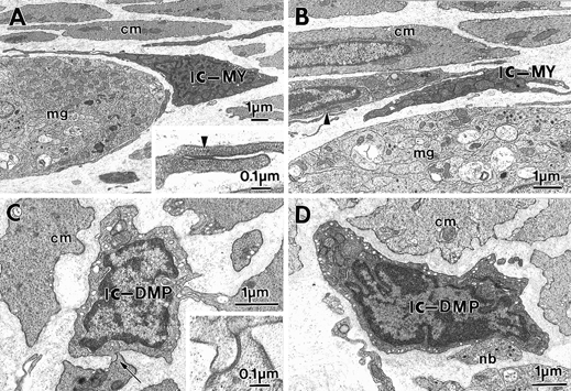
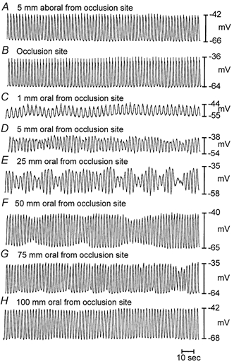
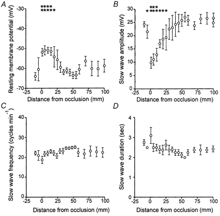
1. Partial obstruction of the murine ileum led to changes in the gross morphology and ultrastructure of the tunica muscularis. Populations of interstitial cells of Cajal (ICC) decreased oral, but not aboral, to the site of obstruction. Since ICC generate and propagate electrical slow waves in gastrointestinal muscles, we investigated whether the loss of ICC leads to loss of function in partial bowel obstruction. 2. Changes in ICC networks and electrical activity were monitored in the obstructed murine intestine using immunohistochemistry, electron microscopy and intracellular electrophysiological techniques. 3. Two weeks following the onset of a partial obstruction, the bowel increased in diameter and hypertrophy of the tunica muscularis was observed oral to the obstruction site. ICC networks were disrupted oral to the obstruction, and this disruption was accompanied by the loss of electrical slow waves and responses to enteric nerve stimulation. These defects were not observed aboral to the obstruction. 4. Ultrastructural analysis revealed no evidence of cell death in regions where the lesion in ICC networks was developing. Cells with a morphology intermediate between smooth muscle cells and fibroblasts were found in locations that are typically populated by ICC. These cells may have been the redifferentiated remnants of ICC networks. 5. Removal of the obstruction led to the redevelopment of ICC networks and recovery of slow wave activity within 30 days. Neural responses were partially restored in 30 days. 6. These data describe the plasticity of ICC networks in response to partial obstruction. After obstruction the ICC phenotype was lost, but these cells regenerated when the obstruction was removed. This model may be an important tool for evaluating the cellular/molecular factors responsible for the regulation and maintenance of the ICC phenotype.
Figures
Similar articles
-
Development of electrical rhythmicity in the murine gastrointestinal tract is specifically encoded in the tunica muscularis.J Physiol. 1997 Nov 15;505 ( Pt 1)(Pt 1):241-58. doi: 10.1111/j.1469-7793.1997.241bc.x. J Physiol. 1997. PMID: 9409486 Free PMC article.
-
Down-regulation of hydrogen sulfide biosynthesis accompanies murine interstitial cells of Cajal dysfunction in partial ileal obstruction.PLoS One. 2012;7(11):e48249. doi: 10.1371/journal.pone.0048249. Epub 2012 Nov 1. PLoS One. 2012. PMID: 23133623 Free PMC article.
-
Interstitial cells of cajal generate electrical slow waves in the murine stomach.J Physiol. 1999 Jul 1;518(Pt 1):257-69. doi: 10.1111/j.1469-7793.1999.0257r.x. J Physiol. 1999. PMID: 10373707 Free PMC article.
-
Physiology and pathophysiology of the interstitial cell of Cajal: from bench to bedside. I. Functional development and plasticity of interstitial cells of Cajal networks.Am J Physiol Gastrointest Liver Physiol. 2001 Sep;281(3):G602-11. doi: 10.1152/ajpgi.2001.281.3.G602. Am J Physiol Gastrointest Liver Physiol. 2001. PMID: 11518672 Review.
-
The interstitial cells of Cajal and a gastroenteric pacemaker system.Arch Histol Cytol. 2002 Mar;65(1):1-26. doi: 10.1679/aohc.65.1. Arch Histol Cytol. 2002. PMID: 12002607 Review.
Cited by
-
Inactivation of inducible nitric oxide synthase protects intestinal pacemaker cells from postoperative damage.J Physiol. 2007 Jul 15;582(Pt 2):755-65. doi: 10.1113/jphysiol.2006.126482. Epub 2007 May 17. J Physiol. 2007. PMID: 17510193 Free PMC article.
-
Voltage dependent potassium channel remodeling in murine intestinal smooth muscle hypertrophy induced by partial obstruction.PLoS One. 2014 Feb 6;9(2):e86109. doi: 10.1371/journal.pone.0086109. eCollection 2014. PLoS One. 2014. PMID: 24516526 Free PMC article.
-
Roles of interleukin-9 in the growth and cholecystokinin-induced intracellular calcium signaling of cultured interstitial cells of Cajal.PLoS One. 2014 Apr 22;9(4):e95898. doi: 10.1371/journal.pone.0095898. eCollection 2014. PLoS One. 2014. PMID: 24755995 Free PMC article.
-
Biomechanical remodelling of obstructed guinea pig jejunum.J Biomech. 2010 May 7;43(7):1322-9. doi: 10.1016/j.jbiomech.2010.01.018. Epub 2010 Mar 1. J Biomech. 2010. PMID: 20189575 Free PMC article.
-
Understanding the Biology of Human Interstitial Cells of Cajal in Gastrointestinal Motility.Int J Mol Sci. 2020 Jun 25;21(12):4540. doi: 10.3390/ijms21124540. Int J Mol Sci. 2020. PMID: 32630607 Free PMC article. Review.
References
-
- Faussone-Pellegrini MS, Cortesini C. The muscle coat of the lower esophageal sphincter in patients with achalasia and hypertensive sphincter. An electron microscopic study. Journal of Submicroscopic Cytology. 1985;17:673–685. - PubMed
-
- Friesen DL, Henderson RD, Hanna W. Ultrastructure of the esophageal muscle in achalasia and diffuse esophageal spasm. American Journal of Clinical Pathology. 1983;79:319–325. - PubMed
-
- Gabella G. Hypertrophic smooth muscle. I. Size and shape of cells, occurrence of mitoses. II Sarcoplasmic reticulum, caveolae and mitochondria. Cell and Tissue Research. 1979;201:63–92. - PubMed
Publication types
MeSH terms
Substances
Grants and funding
LinkOut - more resources
Full Text Sources
Medical