

The 1.19 A X-ray structure of 2'-O-Me(CGCGCG)(2) duplex shows dehydrated RNA with 2-methyl-2,4-pentanediol in the minor groove
- PMID: 11600703
- PMCID: PMC60216
- DOI: 10.1093/nar/29.20.4144
The 1.19 A X-ray structure of 2'-O-Me(CGCGCG)(2) duplex shows dehydrated RNA with 2-methyl-2,4-pentanediol in the minor groove
Abstract
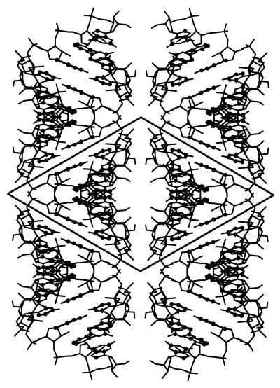
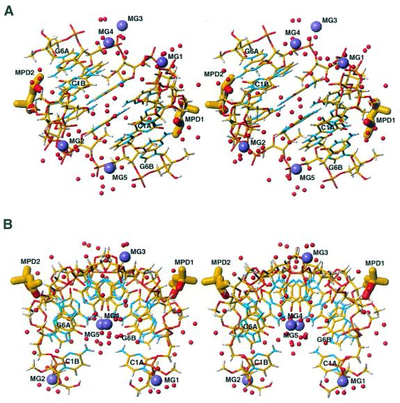
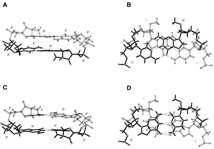
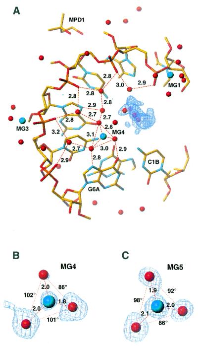
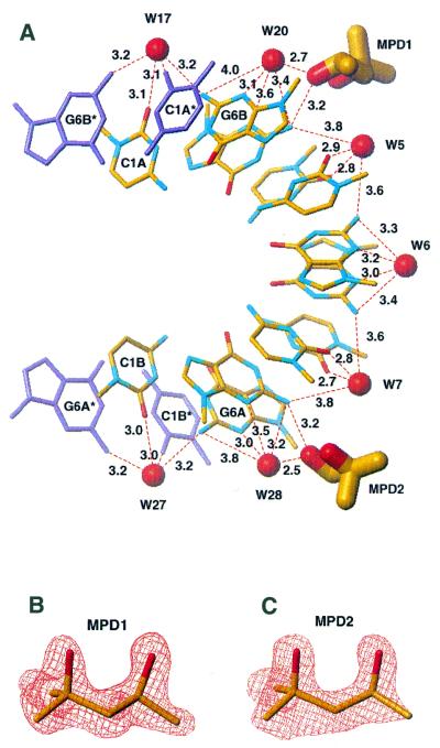
The crystal and molecular structure of 2'-O-Me(CGCGCG)(2) has been determined at 1.19 A resolution, at 100 K, using synchrotron radiation. The structure in space group P3(2)12 is a half-turn right-handed helix that includes two 2-methyl-2,4-pentanediol (MPD) molecules bound in the minor groove. The structure deviates from A-form RNA. The duplex is overwound with an average value of 9.7 bp per turn, characterised as having a C3'-endo sugar pucker, very low base pair rise and high helical twist and inclination angles. The structure includes 65 ordered water molecules. Only a single row of water molecules is observed in the minor groove due to the presence of hydrophobic 2'-O-methyl groups. As many as five magnesium ions are located in the structure. Two are in the major groove and interact with O(6) and N(7) of guanosine and N(4) of cytidine residues through their hydration spheres. This work provides the first example of molecular interactions of nucleic acids with MPD, which was used as a precipitant, cryo-solvent and resolution enhancing agent. The two MPD molecules intrude into the hydration network in the minor groove, each forming hydrogen bonds between their secondary hydroxyl group and exo-amino functions of guanosine residues. Comparison of the 2'-O-Me(CGCGCG)(2) structure in the P3(2)12 and P6(1)22 crystals delineates stability of the water network within the minor groove to dehydration by MPD and is of interest for evaluating factors governing small molecule binding to RNA. Intrusion of MPD into the minor groove of 2'-O-Me(CGCGCG)(2) is discussed with respect to RNA dehydration, a prerequisite of Z-RNA formation.
Figures
References
-
- Arnott S., Hukins,D.W.L. and Dover,S.D. (1972) Optimised parameters for RNA double-helices. Biochem. Biophys. Res. Commun., 48, 1392–1399. - PubMed
-
- Saenger W. (1984) Principles of Nucleic Acids Structure. Springer, Berlin, Germany.
-
- Berman H.M., Zardecki,C. and Westbrook,J. (1998) The Nucleic Acid Database: A resource for nucleic acid science. Acta Crystallogr., 54D, 1095–1104. - PubMed
-
- Auffinger P. and Westhof,E. (1998) Hydration of RNA base pairs. J. Biomol. Struct. Dyn., 16, 693–707. - PubMed
-
- Cate J.H., Gooding,A.R., Podell,E., Zhou,K., Golden,B.L., Kundrot,C.E., Cech,T.R. and Doudna,J.A. (1996) Crystal structure of a group I ribozyme domain: principles of RNA packing. Science, 273, 1678–1685. - PubMed
Publication types
MeSH terms
Substances
Associated data
- Actions
LinkOut - more resources
Full Text Sources
Other Literature Sources
Research Materials
Miscellaneous
