. 2001 Oct 15;29(20):4154-65.
doi: 10.1093/nar/29.20.4154.

Highly conserved amino acids in Pax and Ets proteins are required for DNA binding and ternary complex assembly

Affiliations

Highly conserved amino acids in Pax and Ets proteins are required for DNA binding and ternary complex assembly

D Fitzsimmons et al. Nucleic Acids Res. .

Abstract

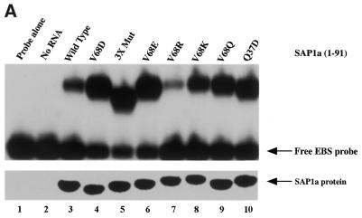
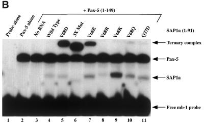
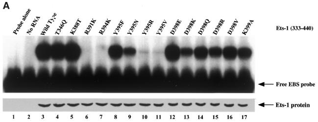
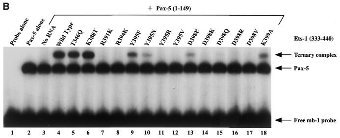
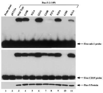
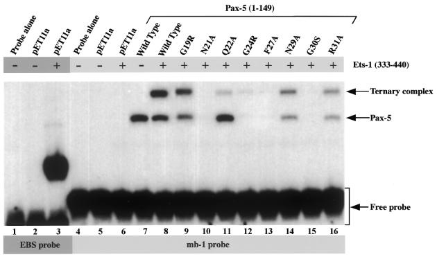
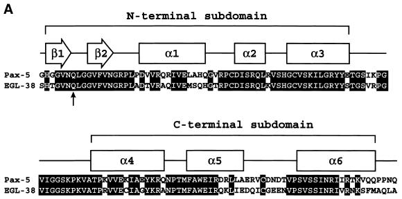
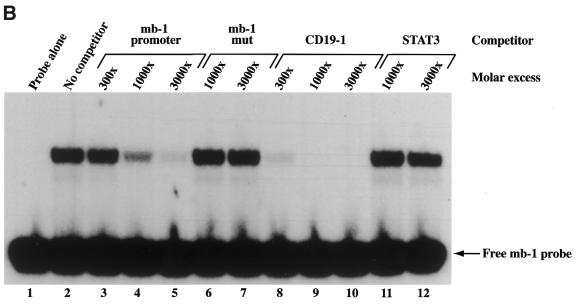
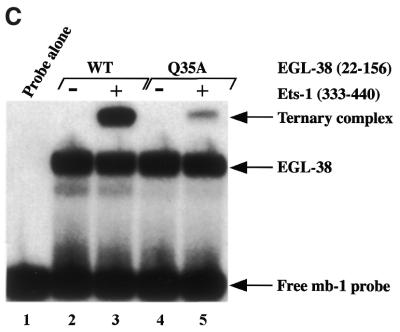
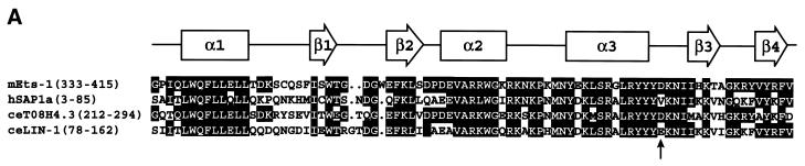
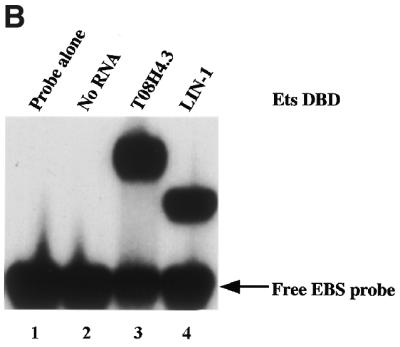
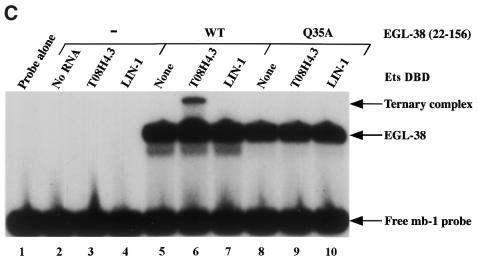
Combinatorial association of DNA-binding proteins on composite binding sites enhances their nucleotide sequence specificity and functional synergy. As a paradigm for these interactions, Pax-5 (BSAP) assembles ternary complexes with Ets proteins on the B cell-specific mb-1 promoter through interactions between their respective DNA-binding domains. Pax-5 recruits Ets-1 to bind the promoter, but not the closely related Ets protein SAP1a. Here we show that, while several different mutations increase binding of SAP1a to an optimized Ets binding site, only conversion of Val68 to an acidic amino acid facilitates ternary complex assembly with Pax-5 on the mb-1 promoter. This suggests that enhanced DNA binding by SAP1a is not sufficient for recruitment by Pax-5, but instead involves protein-protein interactions mediated by the acidic side chain. Recruitment of Ets proteins by Pax-5 requires Gln22 within the N-terminal beta-hairpin motif of its paired domain. The beta-hairpin also participates in recognition of a subset of Pax-5-binding sites. Thus, Pax-5 incorporates protein-protein interaction and DNA recognition functions in a single motif. The Caenorhabditis elegans Pax protein EGL-38 also binds specifically to the mb-1 promoter and recruits murine Ets-1 or the C.elegans Ets protein T08H4.3, but not the related LIN-1 protein. Together, our results define specific amino acid requirements for Pax-Ets ternary complex assembly and show that the mechanism is conserved between evolutionarily related proteins of diverse animal species. Moreover, the data suggest that interactions between Pax and Ets proteins are an important mechanism that regulates fundamental biological processes in worms and humans.


Figures

Figure 1
Figure 1
DNA binding by wild-type and mutated SAP1a ETS domain proteins in the absence or presence of Pax-5. (A) Binding of SAP1a(1–91) polypeptides to the optimized Ets-1 binding site (EBS) probe sequence (5′-CCGGAAG). (Top) EMSA of SAP1a binding to the EBS probe. Synthetic RNAs were translated in wheatgerm extracts in vitro. An aliquot of 1 µl of each programmed extract was incubated with 32P-labeled EBS probe for EMSA as described (24). 3X Mut, K17E/P18Q/V68D. (Bottom) An aliquot of 5 µl of each 35S-labeled translation reaction was fractionated by 18% SDS–PAGE, dried and quantitated on a Molecular Dynamics PhosphorImager. (B) Assembly of Pax-5–SAP1a ternary complexes. In vitro translated SAP1a ETS domain proteins were incubated with the Pax-5 paired domain (1–149) as shown for analysis of binding to 32P-labeled mb-1 probe. No RNA, unprogrammed wheatgerm extract. Bands representing SAP1a alone, Pax-5 alone or Pax-5–SAP1a ternary complexes are indicated on the right.
Figure 1
Figure 1
DNA binding by wild-type and mutated SAP1a ETS domain proteins in the absence or presence of Pax-5. (A) Binding of SAP1a(1–91) polypeptides to the optimized Ets-1 binding site (EBS) probe sequence (5′-CCGGAAG). (Top) EMSA of SAP1a binding to the EBS probe. Synthetic RNAs were translated in wheatgerm extracts in vitro. An aliquot of 1 µl of each programmed extract was incubated with 32P-labeled EBS probe for EMSA as described (24). 3X Mut, K17E/P18Q/V68D. (Bottom) An aliquot of 5 µl of each 35S-labeled translation reaction was fractionated by 18% SDS–PAGE, dried and quantitated on a Molecular Dynamics PhosphorImager. (B) Assembly of Pax-5–SAP1a ternary complexes. In vitro translated SAP1a ETS domain proteins were incubated with the Pax-5 paired domain (1–149) as shown for analysis of binding to 32P-labeled mb-1 probe. No RNA, unprogrammed wheatgerm extract. Bands representing SAP1a alone, Pax-5 alone or Pax-5–SAP1a ternary complexes are indicated on the right.
Figure 2
Figure 2
DNA binding by wild-type and mutated Ets-1 ETS domain proteins in the absence or presence of Pax-5. (A) Binding of Ets-1(333–440) polypeptides to the optimized EBS probe. (Top) EMSA of Ets-1 binding to the EBS probe. Synthetic RNAs were translated in reticulocyte lystates in vitro. An aliquot of 1 µl of each programmed extract was incubated with 32P-labeled EBS probe for EMSA as described (24). (Bottom) An aliquot of 5 µl of each 35S-labeled translation reaction was fractionated by 18% SDS–PAGE, dried and quantitated on a Molecular Dynamics PhosphorImager. (B) Assembly of Pax-5–Ets-1 ternary complexes. In vitro translated SAP1a ETS domain proteins were incubated with Pax-5 paired domain (1–149) as shown for analysis of binding to 32P-labeled mb-1 probe. No RNA, unprogrammed reticulocyte lysate. Bands representing Pax-5 alone or Pax-5–Ets-1 ternary complexes are indicated on the right.
Figure 2
Figure 2
DNA binding by wild-type and mutated Ets-1 ETS domain proteins in the absence or presence of Pax-5. (A) Binding of Ets-1(333–440) polypeptides to the optimized EBS probe. (Top) EMSA of Ets-1 binding to the EBS probe. Synthetic RNAs were translated in reticulocyte lystates in vitro. An aliquot of 1 µl of each programmed extract was incubated with 32P-labeled EBS probe for EMSA as described (24). (Bottom) An aliquot of 5 µl of each 35S-labeled translation reaction was fractionated by 18% SDS–PAGE, dried and quantitated on a Molecular Dynamics PhosphorImager. (B) Assembly of Pax-5–Ets-1 ternary complexes. In vitro translated SAP1a ETS domain proteins were incubated with Pax-5 paired domain (1–149) as shown for analysis of binding to 32P-labeled mb-1 probe. No RNA, unprogrammed reticulocyte lysate. Bands representing Pax-5 alone or Pax-5–Ets-1 ternary complexes are indicated on the right.
Figure 3
Figure 3
Relative binding of wild-type or mutated Pax-5 paired domain (1–149) polypeptides to mb-1 or CD19 probe DNAs. Proteins were synthesized in E.coli as described in Materials and Methods. (Top) EMSA showing binding of Pax-5 to the mb-1 probe. (Center) EMSA showing binding of Pax-5 to the CD19 probe. (Botttom) Relative expression of Pax-5 paired domain proteins in E.coli extracts was determined by analysis of 5 µg total bacterial protein using 18% SDS–PAGE, staining with Coomassie Brilliant Blue and comparing concentrations using a Nucleovision Imaging Workstation (Nucleotech, San Carlos, CA).
Figure 4
Figure 4
Recruitment of the recombinant Ets-1 ETS domain by wild-type or mutated Pax-5 paired domain proteins. Control bacterial lysate generated from E.coli containing empty expression vector (pET11a) or lysates containing bacterially expressed wild-type or mutant Pax-5(1–149) proteins were added to binding reactions as indicated above and analyzed using EMSA. Addition of in vitro translated wild-type Ets-1 ETS domain (333–440) is indicated by + or – above. Binding of Ets-1 alone to the EBS probe (lanes 1–3) or with Pax-5 to the mb-1 probe (lanes 4–16) is indicated below. Bands comprising Pax-5 alone or Pax-5–Ets-1 ternary complexes are indicated on the right.
Figure 5
Figure 5
Caenrhabditis elegans Pax protein EGL-38 binds mb-1 promoter DNA specifically and recruits murine Ets-1. (A) Alignment of amino acid sequences of Pax-5(16–149) versus EGL-38(29–162). The paired domain of Pax-5 is defined as amino acids 16–143 (1). The predicted secondary structure of human Pax-6 is indicated above for reference (3). Boxes, α-helical regions; arrows, β-sheet. Approximate boundaries of the N- and C-terminal subdomains are highlighted. The filled arrow indicates the position of alignment of Gln22 of Pax-5 and Gln35 of EGL-38. (B) Competitive EMSA with the EGL-38 paired domain (22–156). Increasing amounts of double-stranded oligonucleotide competitor DNAs were incubated as indicated above with bacterially expressed EGL-38 and the 32P-labeled mb-1 probe prior to fractionation on a non-denaturing polyacrylamide gel. (C) EGL-38 recruits murine Ets-1 to bind the mb-1 promoter. EMSA was performed using the mb-1 probe with EGL-38 and recombinant murine Ets-1(333–440) as indicated.
Figure 5
Figure 5
Caenrhabditis elegans Pax protein EGL-38 binds mb-1 promoter DNA specifically and recruits murine Ets-1. (A) Alignment of amino acid sequences of Pax-5(16–149) versus EGL-38(29–162). The paired domain of Pax-5 is defined as amino acids 16–143 (1). The predicted secondary structure of human Pax-6 is indicated above for reference (3). Boxes, α-helical regions; arrows, β-sheet. Approximate boundaries of the N- and C-terminal subdomains are highlighted. The filled arrow indicates the position of alignment of Gln22 of Pax-5 and Gln35 of EGL-38. (B) Competitive EMSA with the EGL-38 paired domain (22–156). Increasing amounts of double-stranded oligonucleotide competitor DNAs were incubated as indicated above with bacterially expressed EGL-38 and the 32P-labeled mb-1 probe prior to fractionation on a non-denaturing polyacrylamide gel. (C) EGL-38 recruits murine Ets-1 to bind the mb-1 promoter. EMSA was performed using the mb-1 probe with EGL-38 and recombinant murine Ets-1(333–440) as indicated.
Figure 6
Figure 6
Recruitment of C.elegans Ets proteins by EGL-38. (A) Alignment of the amino acid sequences of the ETS domains of murine Ets-1, human SAP1a, C.elegans LIN-1 and C.elegans T08H4.3. Predicted secondary structure of murine Ets-1 is indicated above (22). Boxes, α-helical regions; arrows, β-sheet. The filled arrow indicates alignment of amino acids with Asp398 of Ets-1. (B) Binding of C.elegans Ets proteins to the EBS probe. EMSA was performed with EBS probe and Ets proteins synthesized in reticulocyte lysates. (C) Recruitment of the T08H4.3 ETS domain, but not that of LIN-1, by the EGL-38 paired domain to bind the mb-1 probe. Recombinant proteins were incubated with mb-1 probe as shown for analysis using EMSA. Bands including EGL-38 alone or ternary complexes with Ets proteins are indicated on the right.
Figure 6
Figure 6
Recruitment of C.elegans Ets proteins by EGL-38. (A) Alignment of the amino acid sequences of the ETS domains of murine Ets-1, human SAP1a, C.elegans LIN-1 and C.elegans T08H4.3. Predicted secondary structure of murine Ets-1 is indicated above (22). Boxes, α-helical regions; arrows, β-sheet. The filled arrow indicates alignment of amino acids with Asp398 of Ets-1. (B) Binding of C.elegans Ets proteins to the EBS probe. EMSA was performed with EBS probe and Ets proteins synthesized in reticulocyte lysates. (C) Recruitment of the T08H4.3 ETS domain, but not that of LIN-1, by the EGL-38 paired domain to bind the mb-1 probe. Recombinant proteins were incubated with mb-1 probe as shown for analysis using EMSA. Bands including EGL-38 alone or ternary complexes with Ets proteins are indicated on the right.

Similar articles

Cited by

References

    1. Czerny T., Schaffner,G. and Busslinger,M. (1993) DNA sequence recognition by Pax proteins: bipartite structure of the paired domain and its binding site. Genes Dev., 7, 2048–2061. - PubMed
    1. Xu W., Rould,M.A., Jun,S., Desplan,C. and Pabo,C.O. (1995) Crystal structure of a paired domain-DNA complex at 2.5 Å resolution reveals structural basis for Pax developmental mutations. Cell, 80, 639–650. - PubMed
    1. Xu H.E., Rould,M.A., Xu,W., Epstein,J.A., Maas,R.L. and Pabo,C.O. (1999) Crystal structure of the human Pax6 paired domain-DNA complex reveals specific roles for the linker region and carboxy-terminal subdomain in DNA binding. Genes Dev., 13, 1263–1275. - PMC - PubMed
    1. Dörfler P. and Busslinger,M. (1996) C-terminal activating and inhibitory domains determine the transactivation potential of BSAP (Pax-5), Pax-2 and Pax-8. EMBO J., 15, 1971–1982. - PMC - PubMed
    1. Eberhard D., Jiménez,G., Heavey,B. and Busslinger,M. (2000) Transcriptional repression by Pax5 (BSAP) through interaction with corepressors of the Groucho family. EMBO J., 19, 2292–2303. - PMC - PubMed

Publication types

MeSH terms