

Degeneration of a homing endonuclease and its target sequence in a wild yeast strain
- PMID: 11600710
- PMCID: PMC60219
- DOI: 10.1093/nar/29.20.4215
Degeneration of a homing endonuclease and its target sequence in a wild yeast strain
Abstract
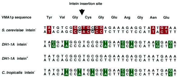
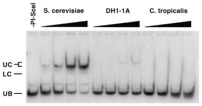
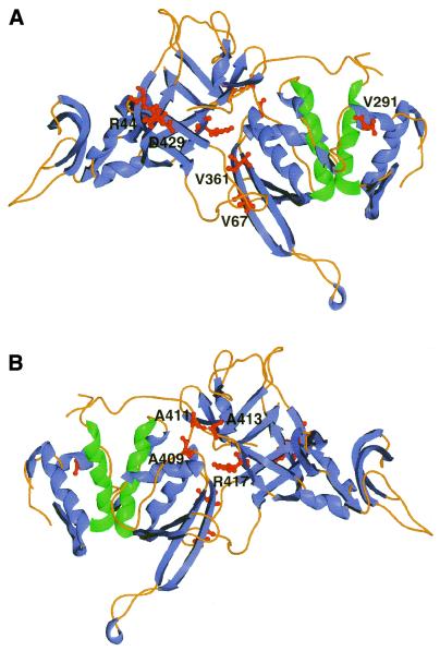
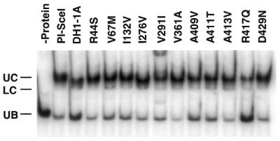
Mobile introns and inteins self-propagate by 'homing', a gene conversion process initiated by site-specific homing endonucleases. The VMA intein, which encodes the PI-SceI endonuclease in Saccharomyces cerevisiae, is present in several different yeast strains. Surprisingly, a wild wine yeast (DH1-1A) contains not only the intein(+) allele, but also an inteinless allele that has not undergone gene conversion. To elucidate how these two alleles co-exist, we characterized the endonuclease encoded by the DH1-1A intein(+) allele and the target site in the intein(-) allele. Sequence analysis reveals seven mutations in the 31 bp recognition sequence, none of which occurs at positions that are individually critical for activity. However, binding and cleavage of the sequence by PI-SceI is reduced 10-fold compared to the S.cerevisiae target. The PI-SceI analog encoded by the DH1-1A intein(+) allele contains 11 mutations at residues in the endonuclease and protein splicing domains. None affects protein splicing, but one, a R417Q substitution, accounts for most of the decrease in DNA cleavage and DNA binding activity of the DH1-1A protein. Loss of activity in the DH1-1A endonuclease and target site provides one explanation for co-existence of the intein(+) and intein(-) alleles.
Figures
Similar articles
-
High resolution crystal structure of domain I of the Saccharomyces cerevisiae homing endonuclease PI-SceI.Nucleic Acids Res. 2002 Sep 15;30(18):3962-71. doi: 10.1093/nar/gkf523. Nucleic Acids Res. 2002. PMID: 12235380 Free PMC article.
-
Substrate recognition and induced DNA distortion by the PI-SceI endonuclease, an enzyme generated by protein splicing.J Mol Biol. 1996 Oct 25;263(2):163-80. doi: 10.1006/jmbi.1996.0567. J Mol Biol. 1996. PMID: 8913299
-
Chimeras of the homing endonuclease PI-SceI and the homologous Candida tropicalis intein: a study to explore the possibility of exchanging DNA-binding modules to obtain highly specific endonucleases with altered specificity.Chembiochem. 2004 Feb 6;5(2):206-13. doi: 10.1002/cbic.200300718. Chembiochem. 2004. PMID: 14760742
-
Homing endonucleases: structural and functional insight into the catalysts of intron/intein mobility.Nucleic Acids Res. 2001 Sep 15;29(18):3757-74. doi: 10.1093/nar/29.18.3757. Nucleic Acids Res. 2001. PMID: 11557808 Free PMC article. Review.
-
Protein splicing: its chemistry and biology.Genes Cells. 1997 Jun;2(6):359-67. doi: 10.1046/j.1365-2443.1997.1270325.x. Genes Cells. 1997. PMID: 9286854 Review.
Cited by
-
Protein Splicing Activity of the Haloferax volcanii PolB-c Intein Is Sensitive to Homing Endonuclease Domain Mutations.Biochemistry. 2020 Sep 15;59(36):3359-3367. doi: 10.1021/acs.biochem.0c00512. Epub 2020 Sep 2. Biochemistry. 2020. PMID: 32822531 Free PMC article.
-
Neighboring inteins interfere with one another's homing capacity.PNAS Nexus. 2023 Oct 27;2(11):pgad354. doi: 10.1093/pnasnexus/pgad354. eCollection 2023 Nov. PNAS Nexus. 2023. PMID: 38024399 Free PMC article.
-
Inteins, introns, and homing endonucleases: recent revelations about the life cycle of parasitic genetic elements.BMC Evol Biol. 2006 Nov 13;6:94. doi: 10.1186/1471-2148-6-94. BMC Evol Biol. 2006. PMID: 17101053 Free PMC article.
-
High resolution crystal structure of domain I of the Saccharomyces cerevisiae homing endonuclease PI-SceI.Nucleic Acids Res. 2002 Sep 15;30(18):3962-71. doi: 10.1093/nar/gkf523. Nucleic Acids Res. 2002. PMID: 12235380 Free PMC article.
-
Mining endonuclease cleavage determinants in genomic sequence data.J Biol Chem. 2011 Sep 16;286(37):32617-27. doi: 10.1074/jbc.M111.259572. Epub 2011 Jul 21. J Biol Chem. 2011. PMID: 21778233 Free PMC article.
References
-
- Kidwell M.G. and Lisch,D.R. (2001) Perspective: transposable elements, parasitic DNA and genome evolution. Evolution, 55, 1–24. - PubMed
-
- Gimble F.S. (2000) Invasion of a multitude of genetic niches by homing endonuclease genes. FEMS Microbiol. Lett., 185, 99–107. - PubMed
-
- Gimble F.S. and Thorner,J. (1992) Homing of a DNA endonuclease gene by meiotic gene conversion in Saccharomyces cerevisiae. Nature, 357, 301–306. - PubMed
Publication types
MeSH terms
Substances
Associated data
- Actions
- Actions
LinkOut - more resources
Full Text Sources
Other Literature Sources
Molecular Biology Databases
Research Materials
Miscellaneous
