. 2001 Oct 15;29(20):E99.
doi: 10.1093/nar/29.20.e99.

A novel yeast system for in vivo selection of recognition sequences: defining an optimal c-Myb-responsive element

Affiliations

A novel yeast system for in vivo selection of recognition sequences: defining an optimal c-Myb-responsive element

T Berge et al. Nucleic Acids Res. .

Abstract

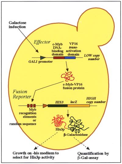
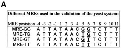
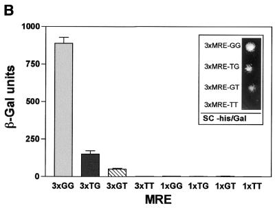
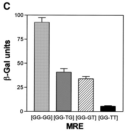
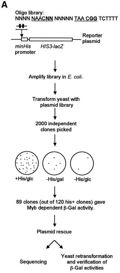
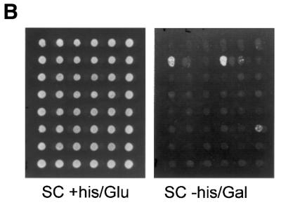
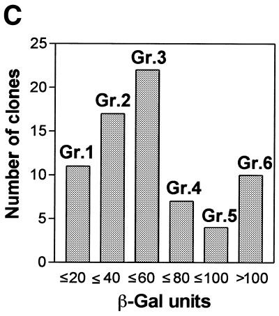
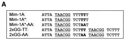
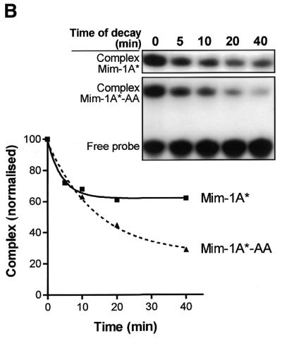
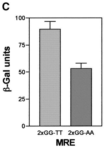
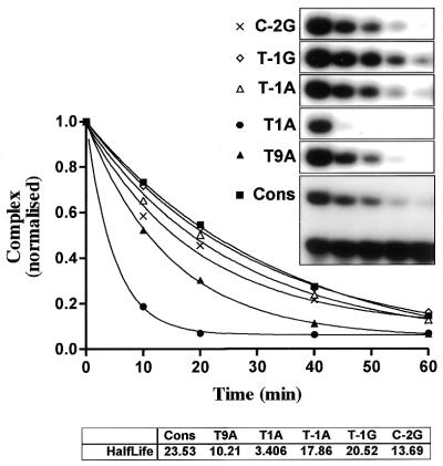
Yeast (Saccharomyces cerevisiae) has proved to be a highly valuable tool in a range of screening methods. We present in this work the design and use of a novel yeast effector-reporter system for selection of sequences recognised by DNA-binding proteins in vivo. A dual HIS3-lacZ reporter under the control of a single randomised response element facilitates both positive growth selection of binding sequences and subsequent quantification of the strength of the selected sequence. A galactose-inducible effector allows discrimination between reporter activation caused by the protein under study and activation due to endogenous factors. The system mimics the physiological gene dosage relationship between transcription factor and target genes in vivo by using a low copy effector plasmid and a high copy reporter plasmid, favouring sequence selectivity. The utility of the novel yeast screening system was demonstrated by using it to refine the definition of an optimal recognition element for the c-Myb transcription factor (MRE). We present screening data supporting an extended MRE consensus closely mimicking known strong response elements and where a sequence of 11 nt influences activity. Novel features include a more strict sequence requirement in the second half-site of the MRE where a T-rich sequence is preferred in vivo.


Figures

Figure 1
Figure 1
The yeast effector–reporter system for in vivo selection of optimal Myb-binding sites. The centromeric effector plasmid pDBD11-R2R3 encodes the chicken c-Myb-R2R3 DNA-binding domain fused to the VP16 transactivation domain and its expression is under control of the GAL1 promoter. The high copy reporter plasmid pYHLfus contains a fusion of two reporter genes, HIS3 and lacZ. A single randomised recognition element allows a simple two-step screening procedure as described in the text.
Figure 2
Figure 2
Validation of the yeast effector–reporter system. (A) To evaluate the system, four different MREs with characterised c-Myb responses (11,24) were inserted in front of the HIS3–lacZ fusion reporter gene. The MREs are designated MRE-GG, MRE-TG, MRE-GT or MRE-TT depending on the nucleotides in MRE positions 5 and 6. After co-transformation of the yeast strain INVSC1 with the Myb effector and one of the different reporter plasmids, growth on medium lacking histidine and β-Gal activity were measured as described in Materials and Methods. (B) Activation of lacZ reporters with single or triple MRE inserts by the chicken c-Myb-R2R3–VP16 effector. (Insert) Activation of HIS3 by chicken c-Myb-R2R3–VP16. Aliquots of 104 cells were spotted on SC galactose medium selective for the plasmids and lacking histidine. (C) Activation of lacZ reporters with inserts containing one constant MRE and one variable MRE (described in the text). The β-Gal assays were performed on exponentially growing cells as described in Materials and Methods. The data in (B) and (C) are presented as mean β-Gal values ± SEM for three independent experiments, each carried out in triplicate.
Figure 2
Figure 2
Validation of the yeast effector–reporter system. (A) To evaluate the system, four different MREs with characterised c-Myb responses (11,24) were inserted in front of the HIS3–lacZ fusion reporter gene. The MREs are designated MRE-GG, MRE-TG, MRE-GT or MRE-TT depending on the nucleotides in MRE positions 5 and 6. After co-transformation of the yeast strain INVSC1 with the Myb effector and one of the different reporter plasmids, growth on medium lacking histidine and β-Gal activity were measured as described in Materials and Methods. (B) Activation of lacZ reporters with single or triple MRE inserts by the chicken c-Myb-R2R3–VP16 effector. (Insert) Activation of HIS3 by chicken c-Myb-R2R3–VP16. Aliquots of 104 cells were spotted on SC galactose medium selective for the plasmids and lacking histidine. (C) Activation of lacZ reporters with inserts containing one constant MRE and one variable MRE (described in the text). The β-Gal assays were performed on exponentially growing cells as described in Materials and Methods. The data in (B) and (C) are presented as mean β-Gal values ± SEM for three independent experiments, each carried out in triplicate.
Figure 3
Figure 3
Screening for optimal MREs in vivo. (A) A schematic view of the screening (see text for details). (B) Clones with a reporter plasmid bearing Myb-binding sites were selected on galactose medium without histidine (SC –His/Gal). An example from 48 independent clones (growing on medium ± histidine) is shown. (C) The β-Gal activities for growth-selected clones were measured as described in the legend to Figure 2 and in Materials and Methods. Based on their β-Gal activity, the clones were divided into six different groups as depicted.
Figure 3
Figure 3
Screening for optimal MREs in vivo. (A) A schematic view of the screening (see text for details). (B) Clones with a reporter plasmid bearing Myb-binding sites were selected on galactose medium without histidine (SC –His/Gal). An example from 48 independent clones (growing on medium ± histidine) is shown. (C) The β-Gal activities for growth-selected clones were measured as described in the legend to Figure 2 and in Materials and Methods. Based on their β-Gal activity, the clones were divided into six different groups as depicted.
Figure 4
Figure 4
Studies of the nucleotides at MRE positions 10 and 11. To determine the importance of the nucleotides at MRE positions 10 and 11 for Myb DNA binding, both in vitro (EMSA) and in vivo (effector–reporter assays in yeast) experiments were performed. (A) The design of the oligos for these studies was based on the mim-1 A-site in the mim-1 promoter (40). The oligos for the EMSA experiments, Mim-1A* and Mim-1A*-AA, contain a C instead of a T in MRE position 8 to prevent the creation of a second MRE in the antiparallel direction. The 2×GG-TT and 2×GG-AA oligos were designed in the same way as the library clones (see Fig. 3A) and were used in the yeast experiments. (B) An EMSA gel showing a time course of decay of the c-Myb-R2R3–DNA complex upon competition with excess non-labelled probe. Myb–DNA complexes were generated using 10 fmol c-Myb-R2R3 and 10 fmol MRE probe. The c-Myb-R2R3–DNA complexes were allowed to form for 15 min at 25°C before they were exposed to a 75-fold excess of non-labelled specific probe for 0, 5, 10, 20 and 40 min. The densities of the complexes were determined, normalised and graphically displayed. (C) Two oligos, 2×GG-TT and 2×GG-AA, were inserted in the pYHLfus reporter plasmid to test Myb-dependent transactivation in yeast. A β-Gal activity assay was performed as described in the legend to Figure 2 and in Materials and Methods. The data are presented as mean β-Gal values ± SEM of three independent experiments, each carried out in triplicate.
Figure 4
Figure 4
Studies of the nucleotides at MRE positions 10 and 11. To determine the importance of the nucleotides at MRE positions 10 and 11 for Myb DNA binding, both in vitro (EMSA) and in vivo (effector–reporter assays in yeast) experiments were performed. (A) The design of the oligos for these studies was based on the mim-1 A-site in the mim-1 promoter (40). The oligos for the EMSA experiments, Mim-1A* and Mim-1A*-AA, contain a C instead of a T in MRE position 8 to prevent the creation of a second MRE in the antiparallel direction. The 2×GG-TT and 2×GG-AA oligos were designed in the same way as the library clones (see Fig. 3A) and were used in the yeast experiments. (B) An EMSA gel showing a time course of decay of the c-Myb-R2R3–DNA complex upon competition with excess non-labelled probe. Myb–DNA complexes were generated using 10 fmol c-Myb-R2R3 and 10 fmol MRE probe. The c-Myb-R2R3–DNA complexes were allowed to form for 15 min at 25°C before they were exposed to a 75-fold excess of non-labelled specific probe for 0, 5, 10, 20 and 40 min. The densities of the complexes were determined, normalised and graphically displayed. (C) Two oligos, 2×GG-TT and 2×GG-AA, were inserted in the pYHLfus reporter plasmid to test Myb-dependent transactivation in yeast. A β-Gal activity assay was performed as described in the legend to Figure 2 and in Materials and Methods. The data are presented as mean β-Gal values ± SEM of three independent experiments, each carried out in triplicate.
Figure 5
Figure 5
Myb–DNA complex dissociation: comparative analysis of variant MREs. Time course of complex dissociation upon competition. The oligos are designated T9A, T1A, T–1A, T–1G and C–2G. They all contain a single base pair deviation from the derived consensus sequence, with the first letter indicating the base specified in the consensus, the number giving the position and the last letter indicating the novel base introduced. Complexes of c-Myb and DNA were generated using 20 fmol c-Myb-R2R3 and 10 fmol different MRE probes (Consensus, T9A, T1A, T–1A, T–1G and C–2G) and allowed to form for 15 min at 25°C with 0.1 µg poly(dI·dC) present. The complexes were then exposed to 750 fmol unlabeled MRE-mim-1A probe for 0, 10, 20, 40 and 60 min before samples were analysed by EMSA as described in Materials and Methods. The densities of the complexes were determined using a phosphorimager, normalised and graphically displayed. The half-lives of the complexes were determined after fitting the points by non-linear regression to a one-phase exponential decay curve.

Similar articles

Cited by

References

    1. Brent R. and Finley,R.L.Jr (1997) Understanding gene and allele function with two-hybrid methods. Annu. Rev. Genet., 31, 663––704.. - PubMed
    1. Fields S. and Song,O. (1989) A novel genetic system to detect protein-protein interactions. Nature, 340, 245––246.. - PubMed
    1. Drees B.L. (1999) Progress and variations in two-hybrid and three-hybrid technologies. Curr. Opin. Chem. Biol., 3, 64––70.. - PubMed
    1. Vidal M. and Legrain,P. (1999) Yeast forward and reverse ‘n’-hybrid systems. Nucleic Acids Res., 27, 919––929.. - PMC - PubMed
    1. Mendelsohn A.R. and Brent,R. (1999) Protein interaction methods—toward an endgame. Science, 284, 1948––1950.. - PubMed

Publication types

Substances