

Review
doi: 10.1136/heart.86.5.579.
Electrophysiology: Ventricular tachycardia: diagnosis of broad QRS complex tachycardia
Affiliations
- PMID: 11602560
- PMCID: PMC1729977
- DOI: 10.1136/heart.86.5.579
Item in Clipboard
Review
Electrophysiology: Ventricular tachycardia: diagnosis of broad QRS complex tachycardia
Heart.
2001 Nov.
No abstract available
Figures
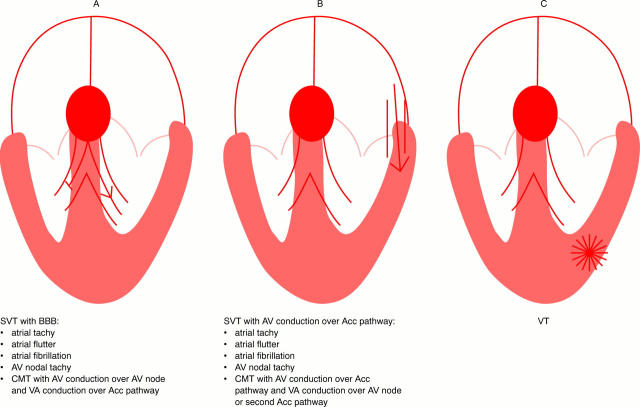
Different types of SVT with BBB (diagram A), SVT with AV conduction over an accessory pathway (diagram B), and VT (diagram C) resulting in a broad QRS tachycardia. Acc, accessory; AV, atrioventricular; BBB, bundle branch block; CMT, circus movement tachycardia; SVT, supraventricular tachycardia; VA, ventriculo-atrial; VT, ventricular tachycardia.
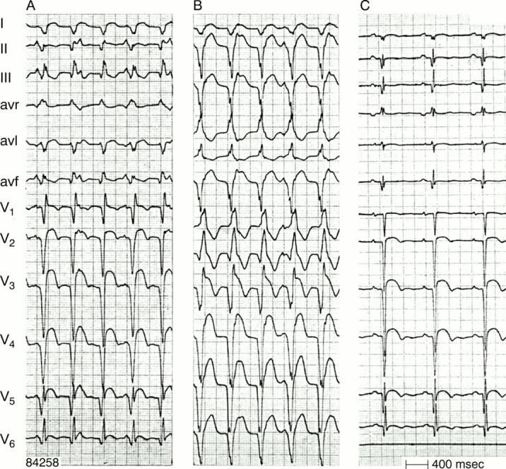
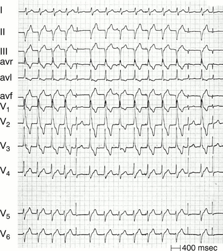
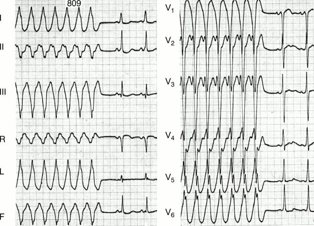
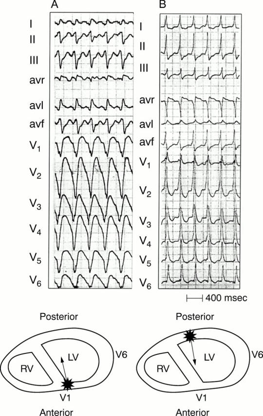
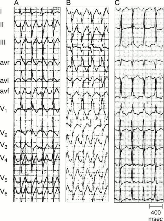
Two types of VT (panel A and B) in the same patient (panel C during sinus rhythm). Atrioventricular dissociation is present during both VTs. Note the effect of the frontal plane axis on the R:S ratio in lead V6 in RBBB shaped VT. R:S < 1 is present in case of a superior axis (panel B), but R:S > 1 with an inferior axis (panel A). Figure 3: One to one ventriculo-atrial conduction during VT. The p waves are negative in leads II, III, and avf and follow each QRS complex. Left panel—VT; right panel—same patient during sinus rhythm.

Two types of VT (panel A and B) in the same patient (panel C during sinus rhythm). Atrioventricular dissociation is present during both VTs. Note the effect of the frontal plane axis on the R:S ratio in lead V6 in RBBB shaped VT. R:S < 1 is present in case of a superior axis (panel B), but R:S > 1 with an inferior axis (panel A). Figure 3: One to one ventriculo-atrial conduction during VT. The p waves are negative in leads II, III, and avf and follow each QRS complex. Left panel—VT; right panel—same patient during sinus rhythm.
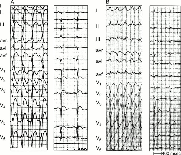
"Capture" (QRS complexes: 5, 13, and 15) and "fusion" beats (QRS complex number 8) during VT.
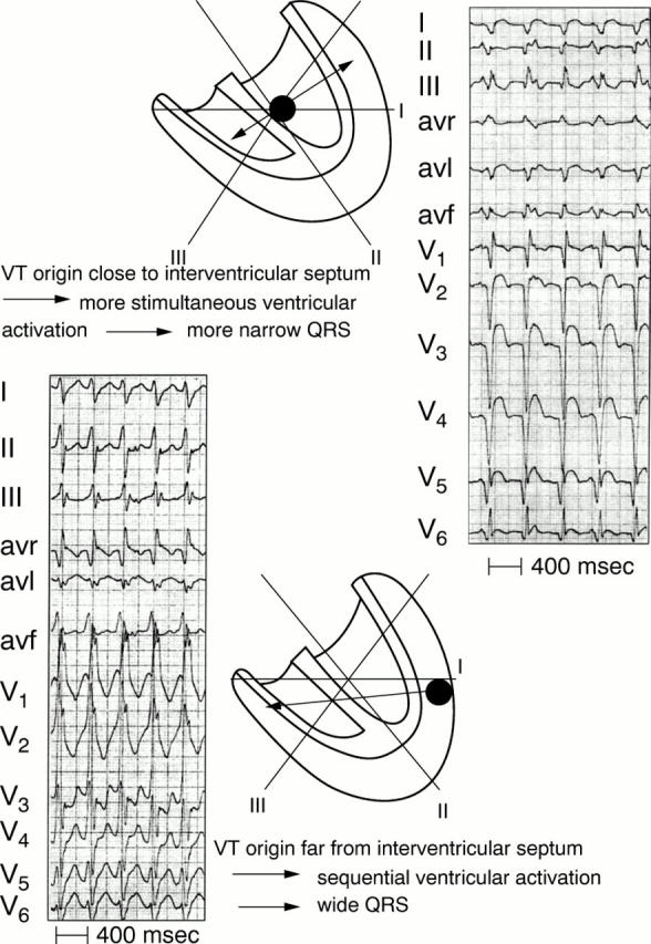
VT origin and QRS width. Upper panel: an origin close to the interventricular septum results in more simultaneous right and left ventricular activation and therefore a more narrow QRS complex. In contrast (lower panel) a VT origin in the lateral ventricular wall results in sequential ventricular activation and a wider QRS complex.
An antidromic circus movement tachycardia with AV conduction over a right sided accessory pathway. The insertion of the accessory pathway in the free wall of the right ventricle results in sequential (right to left) ventricular activation and a wide QRS complex.
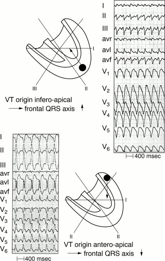
VT origin and QRS axis. An apical origin results in a superiorly directed axis in the frontal plane. In contrast, a basal origin leads to an inferior QRS axis (lower panel).
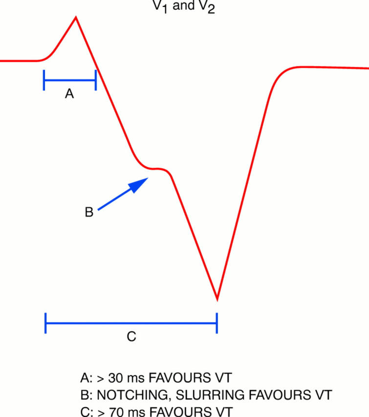
Findings in lead V1 and V2 during LBBB shaped tachycardia pointing to a ventricular origin (see text).
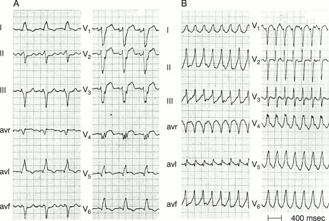
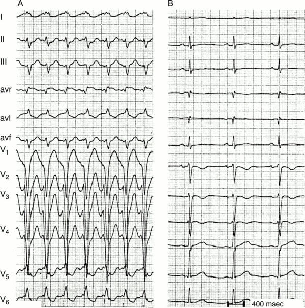
SVT with LBBB. In panel A LBBB changes during tachycardia into a narrow QRS following a ventricular premature beat. As described in the text, lead V1 during LBBB clearly shows signs pointing to a supraventricular origin of the tachycardia. Figure 10: Concordant pattern. The left panel shows a VT arising in the apical area of the left ventricle resulting in negative concordancy of all precordial leads. In the right panel ventricular activation starts in the left posterior area, resulting in positive concordancy of all precordial leads. The latter can be found in left posterior VT but also in SVT with AV conduction over a left posterior accessory pathway.
SVT with LBBB. In panel A LBBB changes during tachycardia into a narrow QRS following a ventricular premature beat. As described in the text, lead V1 during LBBB clearly shows signs pointing to a supraventricular origin of the tachycardia. Figure 10: Concordant pattern. The left panel shows a VT arising in the apical area of the left ventricle resulting in negative concordancy of all precordial leads. In the right panel ventricular activation starts in the left posterior area, resulting in positive concordancy of all precordial leads. The latter can be found in left posterior VT but also in SVT with AV conduction over a left posterior accessory pathway.
Tachycardia QRS smaller than QRS during sinus rhythm. On the left sinus rhythm is present with a very wide QRS because of anterolateral myocardial infarction and pronounced delay in left ventricular activation. On the right a VT arising on the right side of the interventricular septum results in more simultaneous activation of the right and left ventricle than during sinus rhythm and therefore a smaller QRS complex.
QRS complexes during VT indicating a myocardial scar. As shown by the accompanying tracing, during sinus rhythm anterior wall myocardial infarction is present in the left panel and inferior wall myocardial infarction in the right one.
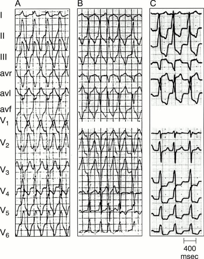
Three types of idiopathic VT arising in or close to the outflow tract of the right ventricle (see text).
Three types of left ventricular idiopathic VT (see text).
VT in arrhythmogenic right ventricular dysplasia (ARVD). VT shows LBBB shape and left axis deviation indicating an origin in the apex of the right ventricle. Note also the negative T waves in V1-V3 during sinus rhythm, which is often found in ARVD.
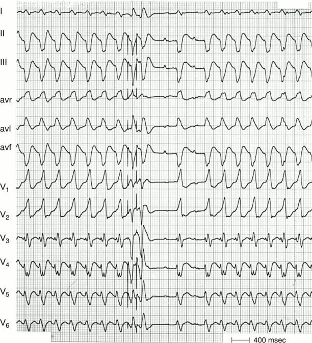
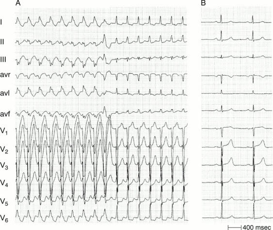
Bundle branch re-entry VT. Following two electrically induced premature beats the tachycardia terminates in the middle of the recording. However, tachycardia resumes after two conducted sinus beats. The QRS is identical during sinus rhythm and tachycardia. Note the presence of AV dissociation during tachycardia indicating a ventricular origin.
References
Publication types
MeSH terms
LinkOut - more resources
Full Text Sources