. 2001 Oct;108(8):1175-83.
doi: 10.1172/JCI12204.

Antigenicity and immunogenicity of allogeneic retinal transplants

Affiliations

Antigenicity and immunogenicity of allogeneic retinal transplants

N G Anosova et al. J Clin Invest. 2001 Oct.

Abstract

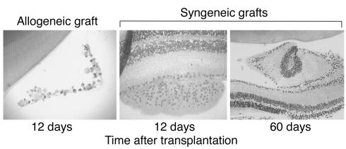
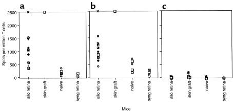
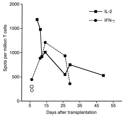
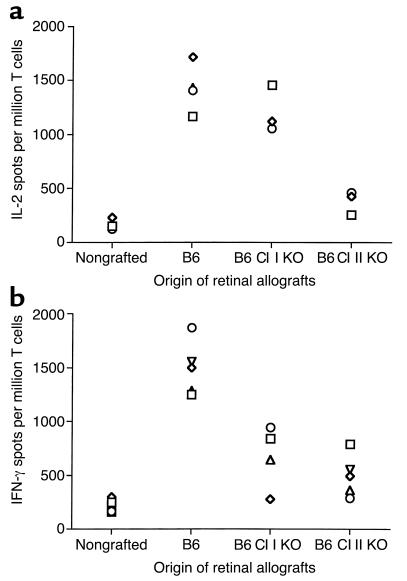
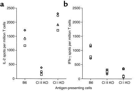
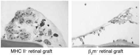
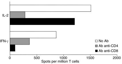
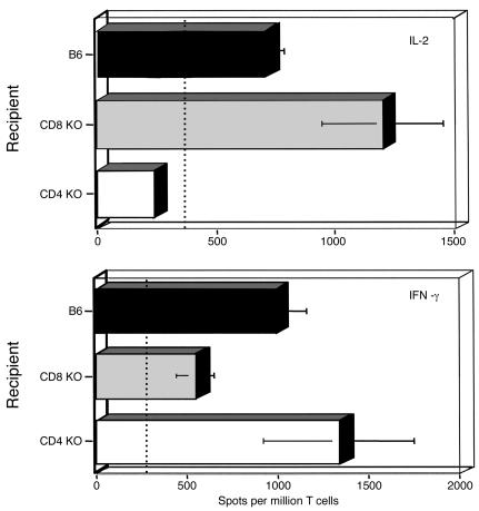
The transplantation of neuronal cells and tissues represents a promising approach for the treatment of incurable neurodegenerative diseases. Indeed, it has been reported recently that retinal transplantation can rescue photoreceptor cells and delay age-related changes in various retinal layers in rodents. However, retinal grafts deteriorate progressively after placement in recipients' eyes. Here we investigated whether a host's immune response elicited toward the graft contributes to its deterioration. Using an ELISA spot assay, we measured T cell responses to retinal tissues placed in the vitreous cavity of syngeneic and allogeneic mice. We found that allogeneic retinas induced potent alloimmune responses mediated by T cells secreting type 1 cytokines (IFN-gamma and IL-2). No response was found in mice engrafted with syngeneic retinas. In addition, all syngeneic retinal grafts displayed no signs of tissue damage (at 55 days), while the majority of allogeneic retinas deteriorated as early as 12 days after placement. Next, we showed that anti-donor responses occurred within two phenotypically and functionally distinct T cell subsets: CD4+ T cells secreting IL-2 and CD8+ T cells producing IFN-gamma. Importantly, CD4+ T cells were necessary and sufficient to cause graft deterioration, while CD8+ T cells did not contribute to this process.


Figures

Figure 1
Figure 1
Histology of retinal transplants placed in the vitreous cavity of recipient mouse eyes. Retinal tissues were stained with H&E. Allogeneic C57BL/6 grafts placed in the eyes of BALB/c mice show a high degree of deterioration (at 12 days after transplantation) (left panel, ×400). In contrast, syngeneic BALB/c retinas placed into BALB/c eyes exhibit well-preserved cell layer architecture both at 12 and 60 days after transplantation (middle and right panels, ×200).
Figure 2
Figure 2
Intravitreal transplantation of allogeneic retinas induces potent immune responses in recipient mice. Recipient BALB/c mice were transplanted with allogeneic (B6) retinas (allo retina), syngeneic retinas (syng retina), or allogeneic skins (skin graft). Nontransplanted mice were used as controls (naive). Ten to nineteen days after transplantation, spleen recipient T cells were restimulated in vitro with irradiated donor splenocytes for 20–24 hours for IL-2 and 40–44 hours for IFN-γ and IL-5. The frequency of (a) IFN-γ–, (b) IL-2–, and (c) IL-5–producing T cells was measured using the ELISA spot technique. The data are expressed as cytokine-producing spots per million T cells. The IFN-γ and IL-2 results obtained after skin grafting (> 2,500 spots/million T cells) are representative of six mice tested individually. In all other cases, each individual symbol corresponds to one mouse.
Figure 3
Figure 3
Kinetics of the immune response in the spleen of retina-transplanted mice. T cells from mice grafted with allogeneic retinas were harvested at different time points after transplantation. T cells were restimulated in vitro with donor irradiated splenocytes. The frequency of IFN-γ–producing (filled circles) and IL-2–producing (filled squares) T cells was determined using ELISA spot methodology. The data are expressed as cytokine-producing spots per million T cells. Open symbols represent the frequency of IFN-γ–producing (open circle) and IL-2–producing (open square) T cells in control naive mice. Each point represents the average number of spots from three to six mice tested individually.
Figure 4
Figure 4
Alloreactive T cell responses in mice grafted with retinas devoid of MHC class I and class II molecules. The number of IL-2–secreting (a) and IFN-γ–secreting (b) T cells was evaluated in recipient BALB/c mice transplanted with retinal grafts from wild-type B6 (positive control), B6 class I KO (β2m–/–; B6 Cl I KO), and B6 Cl II KO donors. T cells from naive, nontransplanted BALB/c mice were used as negative control. In all experiments, T cells were restimulated in vitro with B6 irradiated splenocytes. Each symbol represents an individual mouse. The data are expressed as cytokine-producing spots per million T cells.
Figure 5
Figure 5
Alloreactive responses of T cells from retinal-grafted mice restimulated in vitro with donor APCs lacking MHC class I and class II expression. The number of IL-2–secreting (a) and IFN-γ–secreting (b) T cells was evaluated in BALB/c mice transplanted with retinal grafts from wild-type B6 mice. Recipient T cells were restimulated in vitro with wild-type B6 irradiated cells (positive control) or with donor APCs devoid of either MHC class I or class II expression. The average frequencies of IL-2–producing and IFN-γ–producing cells responding to allogeneic wild-type B6 stimulators in nontransplanted mice was 500 ± 150 and 240 ± 120 spots, n = 15, respectively (negative controls). Each symbol represents an individual mouse. The data are expressed as cytokine-producing spots per million T cells.
Figure 6
Figure 6
Histology of MHC class II and MHC class I KO (β2m KO) allogeneic (B6) retinal grafts placed in the vitreous cavity of BALB/c recipients. Retinal tissues were collected 12 days after transplantation and stained with H&E.
Figure 7
Figure 7
Effects of anti-CD4 and anti-CD8 Ab treatments on alloimmune responses induced after retinal allotransplantation. T cells from retinal-allotransplanted (BALB/c) mice were restimulated in vitro with donor B6 irradiated splenocytes in the presence of anti-CD4 or anti-CD8 mAb’s (2 μg/ml). The frequency of IL-2– and IFN-γ–producing T cells was measured by the ELISA spot method and the results expressed as spots per million T cells. The data shown here are representative of four separate experiments. The data show the frequency of cytokine-producing spots per million T cells.
Figure 8
Figure 8
Alloimmune responses to retinal allografts in recipient mice devoid of CD4 and CD8 T cell subsets. Wild-type (B6) as well as CD4 KO and CD8 KO mice were used as recipients. After intravitreal grafting with allogeneic (BALB/c) retinas, recipient T cells were purified and incubated in vitro with donor irradiated spleen cells. The frequency of activated T cells releasing IL-2 (upper panel) and IFN-γ (lower panel) cytokines was determined by the ELISA spot method. The dotted line represents the frequencies of cytokine-producing T cells obtained in naive B6 mice. The data shown represent the mean number of cytokine-producing spots (per million T cells) ± SD obtained with six to seven mice tested separately.

References

    1. Algvere PV, Berglin L, Gouras P, Sheng Y. Transplantation of fetal retinal pigment epithelium in age-related macular degeneration with subfoveal neovascularization. Graefes Arch Clin Exp Ophthalmol. 1994;232:707–716. - PubMed
    1. del Cerro M, et al. Retinal transplants for cell replacement in phototoxic retinal degeneration. Prog Clin Biol Res. 1989;314:673–686. - PubMed
    1. del Cerro M, et al. Retinal transplants into adult eyes affected by phototoxic retinopathy. Prog Brain Res. 1988;78:125–130. - PubMed
    1. Gouras P, Flood MT, Kjedbye H, Bilek MK, Eggers H. Transplantation of cultured human retinal epithelium to Bruch’s membrane of the owl monkey’s eye. Curr Eye Res. 1985;4:253–265. - PubMed
    1. Gouras P, Lopez R. Transplantation of retinal epithelial cells. Invest Ophthalmol Vis Sci. 1989;30:1681–1683. - PubMed

Publication types

MeSH terms

Substances