Case Reports
. 2001 Oct;108(8):1215-20.
doi: 10.1172/JCI13180.

Familial isolated hypoparathyroidism caused by a mutation in the gene for the transcription factor GCMB

Affiliations
Case Reports

Familial isolated hypoparathyroidism caused by a mutation in the gene for the transcription factor GCMB

C Ding et al. J Clin Invest. 2001 Oct.

Abstract

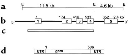
Hypoparathyroidism is characterized by hypocalcemia, hyperphosphatemia, and absent or markedly reduced circulating concentrations of parathyroid hormone. The transcription factor GCMB is predominantly, if not exclusively, expressed in parathyroid cells and is critical for development of the parathyroid glands in mice. Thus, in the present study we examined the GCMB gene, mapped to 6p23-24, as a candidate for isolated hypoparathyroidism. We defined the boundaries of the five exons of the human GCMB gene and then identified a large intragenic mutation in the GCMB genes of the proband of an extensive kindred with isolated hypoparathyroidism. Her parents and several other unaffected relatives were heterozygous for the mutation. Despite an absence of any history of consanguinity, microsatellite analysis showed shared genotypes that flanked the GCMB gene over a span of 5 cM, suggesting that both of the proband's GCMB alleles had been derived from a single common ancestor. Analysis of additional, unrelated cases did not disclose the same mutation. We conclude that homozygous loss of function of the GCMB gene impairs normal parathyroid gland embryology and is responsible for isolated hypoparathyroidism in a subset of patients with this disease.


Figures

Figure 1
Figure 1
Pedigree of family described in text. The figure above shows the pedigree of the family, with the proband indicated by the arrow. Squares denote male family members, circles female family members, and solid symbols subjects with symptomatic hypocalcemia. Markers under each symbol indicate the genotype for the GCMB gene allele; WT denotes wild-type allele; M denotes ΔGCMB allele. NT, not tested.
Figure 2
Figure 2
GCMB gene. (a) An EcoRI restriction map of the human GCMB gene, with approximate genomic distances between restriction sites noted. (b) The genomic organization of the human GCMB gene, with exons 1–5 indicated by the boxes and introns indicated by the connecting lines. The numbers above each exon indicate the corresponding nucleotide number of the cloned cDNA. (c) The area of the gene deleted in the ΔGCMB allele. (d) The cDNA, with the 5′ and 3′ untranslated regions (UTR), and the open reading frame that predicts a protein of 506 amino acids.
Figure 3
Figure 3
Amplification of exons 1 and 2 by PCR. Genomic DNA from four unrelated subjects with familial isolated hypoparathyroidism was amplified using oligonucleotide primers flanking exon 1 (left panel) or exon 2 (right panel); the proband shown in Figure 1 corresponds to subject 2. The positions of molecular-weight markers are indicated on the left. DNA from subjects 1, 3, and 4 showed amplification of appropriate-sized fragments, whereas DNA from subject 2 failed to amplify a fragment corresponding to either exon 1 or exon 2.
Figure 4
Figure 4
Amplification of exons 3, 4, and 5 by PCR. Genomic DNA from the proband (Pt), her father (F) and mother (M), and two unrelated normal subjects (NL) was amplified using oligonucleotide primers flanking exon 3 (upper panel), exon 4 (middle panel), or exon 5 (bottom panel). Arrows indicate the position of the appropriately sized amplified product. DNA from the proband (Pt) failed to amplify fragments corresponding to exons 3 and 4 but did yield an appropriate PCR product corresponding to exon 5. Genomic DNA from the parents and two normal subjects yielded appropriate-sized PCR products for all three exons.
Figure 5
Figure 5
Southern blot analysis of GCMB gene. Genomic DNA from the proband (Pt), her father (F) and mother (M), and two unrelated normal subjects (NL) was digested using EcoRI and hybridized to a radiolabeled genomic DNA corresponding to either exon 3 (left) or exons 3–5 (right). The positions of molecular-weight markers are indicated. Genomic DNA from the proband (Pt) failed to hybridize to the exon 3 probe, and a unique 8.4-kb EcoRI fragment hybridized to the exon 3–5 probe. The proband’s parents showed hybridization of the exon 3–5 probe with two restriction fragments of expected size and with the abnormal 8.4-kb fragment, all with reduced signal intensity (see text).
Figure 6
Figure 6
Haplotype analysis of the GCMB locus. The figure above shows the haplotypes for the GCMB locus obtained using microsatellite polymorphisms. The proband is denoted by the solid circle, and her heterozygous father and mother are indicted by the half-filled square and circle, respectively. This figure demonstrates that the two parental mutant alleles (ΔGCMB) have identical haplotypes for a series of markers that span over 5 cM of genomic DNA on chromosome 6p23–24.

References

    1. Hebert SC, Brown EM. The extracellular calcium receptor. Curr Opin Cell Biol. 1995;7:484–492. - PubMed
    1. Brown EM, et al. Cloning and characterization of an extracellular Ca2+-sensing receptor from bovine parathyroid. Nature. 1993;366:575–580. - PubMed
    1. Chattopadhyay N, Mithal A, Brown EM. The calcium-sensing receptor: a window into the physiology and pathophysiology of mineral ion metabolism. Endocr Rev. 1996;17:289–307. - PubMed
    1. Ahonen P. Autoimmune polyendocrinopathy-candidosis-ectodermal dystrophy (APECED): autosomal recessive inheritance. Clin Genet. 1985;27:535–542. - PubMed
    1. Ahonen P, Myllarniemi S, Sipila I, Perheentupa J. Clinical variation of autoimmune polyendocrinopathy-candidiasis-ectodermal dystrophy (APECED) in a series of 68 patients. N Engl J Med. 1990;322:1829–1836. - PubMed

Publication types