. 2001 Oct 15;194(8):1111-22.
doi: 10.1084/jem.194.8.1111.

A DAP12-mediated pathway regulates expression of CC chemokine receptor 7 and maturation of human dendritic cells

Affiliations

A DAP12-mediated pathway regulates expression of CC chemokine receptor 7 and maturation of human dendritic cells

A Bouchon et al. J Exp Med. .

Abstract

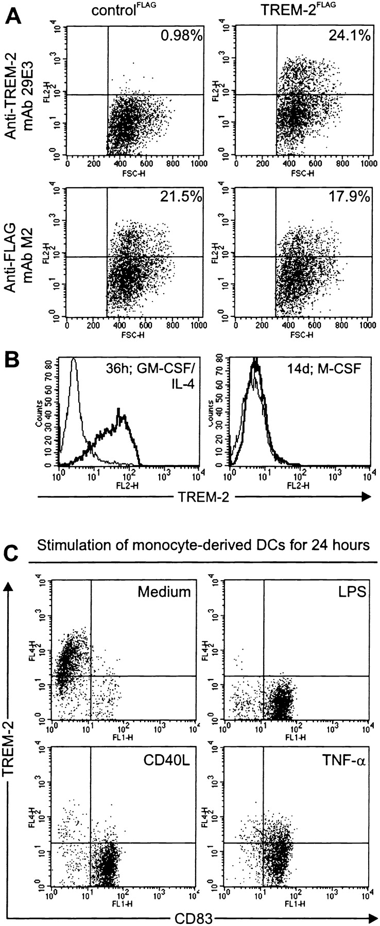
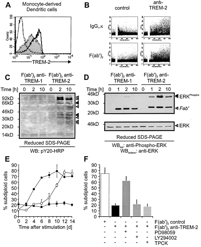
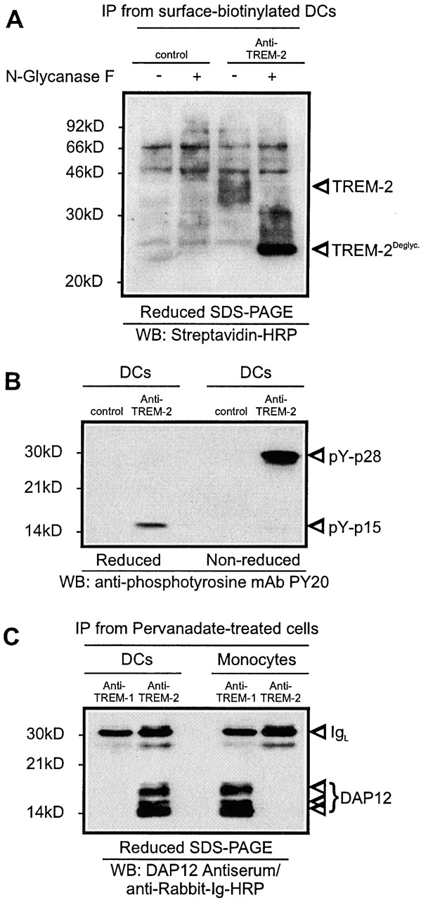
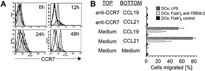
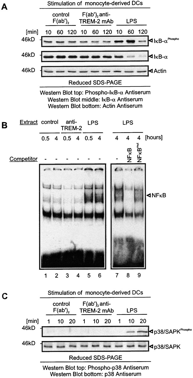
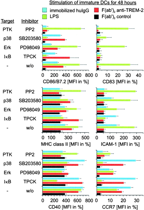
Gene targeting of the adaptor molecule DAP12 in mice caused abnormal distribution and impaired antigen presentation capacity of dendritic cells (DCs). However, the DAP12-associated receptors expressed on DCs and their functions have not been identified yet. Here we show that the triggering receptor expressed on myeloid cells-2 (TREM-2) is a cell surface receptor on human monocyte-derived DCs, which is associated with DAP12. TREM-2/DAP12 promotes upregulation of CC chemokine receptor 7, partial DC maturation, and DC survival through activation of protein tyrosine kinases and extracellular signal-regulated kinase. In contrast to Toll-like receptor-mediated signaling, TREM2/DAP12 stimulation is independent of nuclear factor-kappaB and p38 stress-activated protein kinase. This novel DC activation pathway may regulate DC homeostasis and amplify DC responses to pathogens, explaining the phenotype observed in DAP12-deficient mice.


Figures

Figure 1
Figure 1
TREM-2 is selectively expressed on immature DCs. (A) mAb 29E3 recognizes selectively TREM-2. 293 cells transfected with a cDNA encoding FLAG-tagged TREM-2 (TREM-2FLAG; right panels) were stained with mAb 29E3 (top panel), compared with cells transfected with a control cDNA (controlFLAG; left panels). The percentages of positive cells (top right quadrants) are indicated. Expression of TREM-2FLAG and controlFLAG was confirmed using an anti-FLAG mAb (bottom panels). Cells stained with an isotype-matched control mAbs were comprised within the indicated bottom quadrant. (B) TREM-2 is strongly upregulated after stimulation of monocytes with GM-CSF and IL-4. Monocytes treated with GM-CSF/IL-4 (left panel) or M-CSF (right panel) were analyzed by flow cytometry for cell surface expression of TREM-2 (solid bold line) after 36 h or up to 14 d, respectively. Dashed profiles indicate background staining with a control IgG1 mAb. (C) TREM-2 is rapidly downregulated upon maturation of DCs. LPS- (top right panel), CD40L- (bottom left panel), TNFα-stimulated (bottom right panel), or unstimulated monocyte-derived DCs (top left panel) were analyzed by flow cytometry for cell surface expression of TREM-2 and CD83 after 36 h. Cells stained with an isotype-matched control mAbs were comprised within the indicated bottom quadrants.
Figure 3
Figure 3
Stimulation of monocyte-derived DCs via TREM-2 induces calcium mobilization, tyrosine phosphorylation, and prolongs DC survival by an Erk-dependent pathway. (A) Functional characterization of F(ab′) and F(ab′)2 29E3. Monocyte-derived DCs were analyzed by flow cytometry for cell surface expression of TREM-2 using either biotinylated F(ab′)2 29E3 (solid bold profile), F(ab′) 29E3 (gray profile), control F(ab′) (dashed profile), or F(ab′)2 (solid profile) followed by Streptavidin-PE. (B) Stimulation of TREM-2 induces intracellular Ca2+ mobilization. Bivalent crosslinking of TREM-2 using IgG1, κ 29E3, or Fc-free F(ab′)2 29E3 induces intracellular Ca2+ mobilization in contrast to control mAbs (21C7 anti–TREM-1 mAb; left panels; 1A11 anti-MHC class I mAb; data not shown). (C) Antiphosphotyrosine blot of cell lysates from monocyte-derived DCs stimulated with F(ab′)2 29E3 (anti–TREM-2) or control F(ab′)2 (anti–TREM-1 mAb) for the indicated time periods. (D) Monocyte-derived DCs were stimulated as indicated in C and examined by Western blot analysis for anti-phospho-Erk1/2 (top panel) compared with anti-Erk 1/2 (bottom panel). Arrows indicate phosphorylated proteins in all panels. Molecular weight markers are shown. (E) Monocyte-derived DCs were washed five times to remove GM-CSF/IL-4 before stimulation with plastic-bound F(ab′)2 29E3 (open circles), control F(ab′)2 (21C7 anti–TREM-1 mAb) (filled circles), or GM-CSF (filled squares) for the indicated time periods. Apoptotic cell death was determined by measurement of DNA fragmentation. (F) Monocyte-derived DCs were stimulated plastic-bound F(ab′)2 29E3 in the presence or absence of Erk-inhibition (PD98059 [20 μM]), PI3K-inhibition (LY294002 [10 μM]), or inhibition of IκB-degradation (TPCK [20 μM]). Apoptotic cell death was determined after 8 d compared with DCs stimulated with control F(ab′)2 by measuring subdiploid DNA content.
Figure 2
Figure 2
TREM-1 is a 40-kD glycoprotein associated with the adaptor protein DAP12. (A) Surface-biotinylated monocyte-derived DCs were lysed and subjected to immunoprecipitation with 29E3 anti–TREM-2 mAb (right lanes) or control IgG1 mAbs (21C7 anti–TREM-1 mAb). Immunoprecipitates (IP) were left untreated or treated with N-glycanase F and analyzed by Western blot analysis with Streptavidine-HRP. Molecular weight markers and specific protein bands are indicated. (B) Pervanadate-treated monocyte-derived DCs were subjected to immunoprecipitation with 29E3 anti–TREM-2 mAb or control IgG1 mAb (21C7 anti–TREM-1 mAb). The precipitates were analyzed by antiphosphotyrosine blot under reducing (left lanes) and nonreducing (right lanes) conditions. Tyrosine phosphorylated proteins are marked by arrows. Molecular weight markers are indicated. (C) Anti-DAP12 blot analysis of TREM-2 immunoprecipitate from monocyte-derived DCs (left lanes) and monocytes (right lanes) after pervanadate stimulation (reducing conditions). TREM-1 immunoprecipitates from monocytes and monocyte-derived DCs were included as positive and negative controls, respectively. Molecular weight markers and specific protein bands are indicated.
Figure 4
Figure 4
Stimulation of TREM-2 induces CCR7 expression and chemotactic response toward CCL19 and CCL21. (A) CCR7 is rapidly upregulated after stimulation of TREM-2 on DCs. DCs were stimulated with F(ab′)2 anti–TREM-2 mAb (gray profiles), control F(ab′)2 (21C7; anti–TREM-1; solid line profiles), or LPS (solid bold profiles). After the indicated time periods cells were harvested and analyzed by flow cytometry for cell surface expression of CCR7 by anti-CCR7 mAb (mouse IgM) followed by PE-labeled goat anti–mouse IgM. Dashed profiles indicate background staining with a control IgM mAb. (B) TREM-2 stimulation directs migration of DCs toward CCL19 and CCL21 by a CCR7-dependent pathway. DCs stimulated for 24 h with plastic-coated control F(ab′)2 (21C7, anti–TREM-1; black bars), F(ab′)2 anti–TREM-2 mAb (light gray bars), or LPS (dark gray bars) were tested in Transwell chemotaxis assays for the ability to migrate toward medium alone, medium supplemented with 100 ng/ml CCL19 or CCL21 (BOTTOM well). In control experiments, DCs were preincubated for 15 min with anti-CCR7 mAb before placing them in the TOP well for assessment of chemotaxis toward medium alone, medium supplemented with CCL19, or CCL21 placed in the BOTTOM well.
Figure 5
Figure 5
TREM-2 does not initiate IκBα/NF-κB- and p38/SAPK-dependent pathways. (A) Lack of IκBα phosphorylation and degradation upon TREM-2 stimulation of monocyte-derived DCs. Monocyte-derived DCs were stimulated by LPS or plastic-bound F(ab′)2 anti–TREM-2 mAb or control F(ab′)2 for the indicated times. Protein lysates were tested for IκBα phosphorylation and degradation by Western blot analysis. The same blot was sequentially stripped and reprobed with antiphospho-IκBα, anti-IκBα, and anti-actin (loading control) antibodies. (B) Lack of NF-κB translocation upon TREM-2 triggering. Monocyte-derived DCs were stimulated for the indicated time points as described in panel A and nuclear extracts were obtained. Radiolabeled NF-κB consensus double-stranded oligonucleotides were incubated with the indicated nuclear extracts in the absence of competing oligonucleotides or in the presence of a 25-fold molar excess of wild-type (NF-κB) or mutant (NF-κBmut) competing oligonucleotides. DNA–protein complexes were resolved by electrophoresis. The NF-κB–containing complex is marked. (C) Absence of p38/SAPK phosphorylation upon TREM-2 stimulation of monocyte-derived DCs. Monocyte-derived DCs were stimulated for the indicated times as described in panel A. Protein lysates were tested for p38/SAPK phosphorylation (top panel) by Western blot analysis. The same blot was sequentially stripped and reprobed with anti-p38/SAPK antibodies (bottom panel). Arrows indicate proteins in all panels. Molecular weight markers are shown. To confirm proper stimulation of TREM-2/DAP12 in all experimental settings an aliquot of stimulated DCs was kept and tested after 48 h for upregulation of MHC class II and CD86 (data not shown).
Figure 6
Figure 6
Comparison of TREM-2-, LPS/TLR-, and FcR-mediated maturation pathways. Monocyte-derived DCs were stimulated with plastic-bound control F(ab′)2 (black bars), F(ab′)2 anti–TREM-2 (red bars), human IgG (blue bars), or LPS (green bars) in the presence of inhibitors for Erk (PD98059), p38/SAPK (SB203580), PTK (PP2), IκBα degradation (TPCK), or an equal volume of DMSO as a control. After 48 h, cell surface expression of CD86 (top left panel), MHC class II (middle left panel), CD40 (bottom left panel), CD83 (top right panel), ICAM-1 (middle right panel), or CCR7 (bottom right panel) was determined by flow cytometry. Data shown are representative of four independent experiments and display the mean and standard deviation of three independent samples.

References

    1. Banchereau J., Steinman R.M. Dendritic cells and the control of immunity. Nature. 1998;392:245–252. - PubMed
    1. Gallucci S., Lolkema M., Matzinger P. Natural adjuvantsendogenous activators of dendritic cells. Nat. Med. 1999;11:1249–1255. - PubMed
    1. Sauter B., Albert M.L., Francisco L., Larsson M., Somersan S., Bhardwaj N. Consequences of cell deathexposure to necrotic tumor cells, but not primary tissue cells or apoptotic cells, induces the maturation of immunostimulatory dendritic cells. J. Exp. Med. 2000;191:423–434. - PMC - PubMed
    1. Dieu M.C., Vanbervliet B., Vicari A., Bridon J.M., Oldham E., Ait-Yahia S., Briere F., Zlotnik A., Lebecque S., Caux C. Selective recruitment of immature and mature dendritic cells by distinct chemokines expressed in different anatomic sites. J. Exp. Med. 1998;188:373–386. - PMC - PubMed
    1. Sozzani S., Allavena P., D'Amico G., Luini W., Bianchi G., Kataura M., Imai T., Yoshie O., Bonecchi R., Mantovani A. Differential regulation of chemokine receptors during dendritic cell maturationa model for their trafficking properties. J. Immunol. 1998;161:1083–1086. - PubMed

Publication types

MeSH terms

Substances