. 2001 Oct 15;194(8):1171-8.
doi: 10.1084/jem.194.8.1171.

CD11c(+)B220(+)Gr-1(+) cells in mouse lymph nodes and spleen display characteristics of plasmacytoid dendritic cells

Affiliations

CD11c(+)B220(+)Gr-1(+) cells in mouse lymph nodes and spleen display characteristics of plasmacytoid dendritic cells

H Nakano et al. J Exp Med. .

Abstract

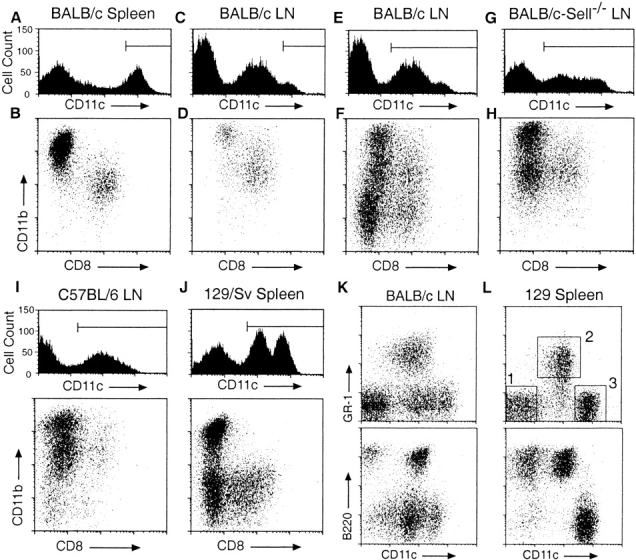
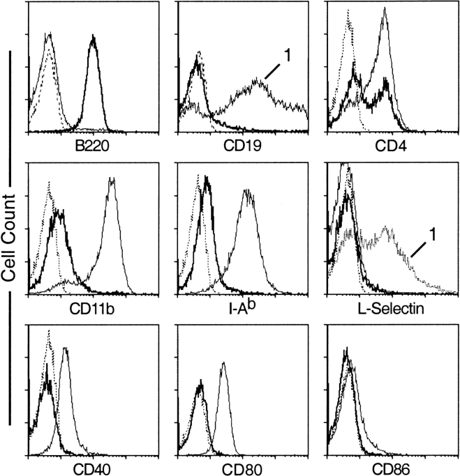
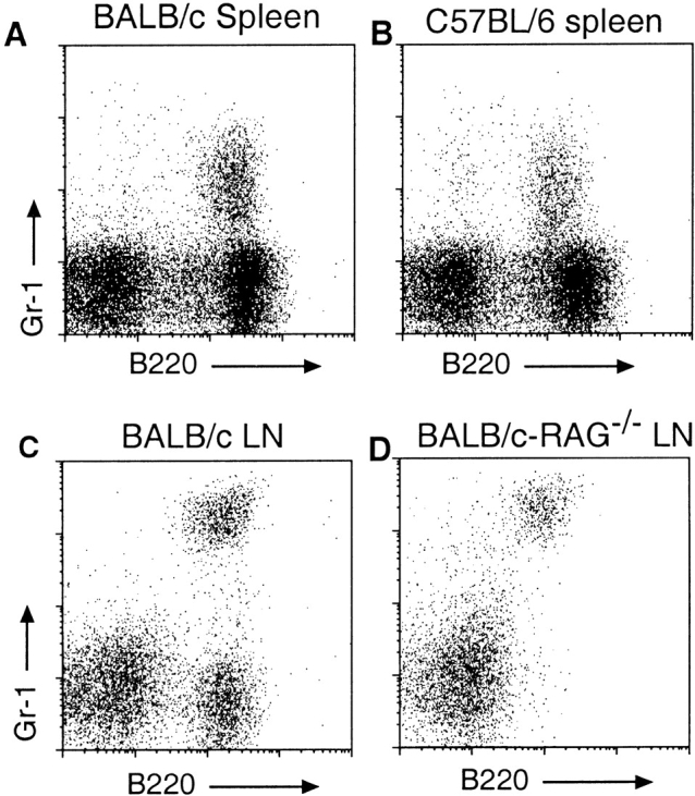
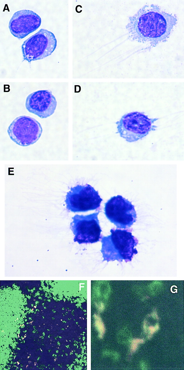
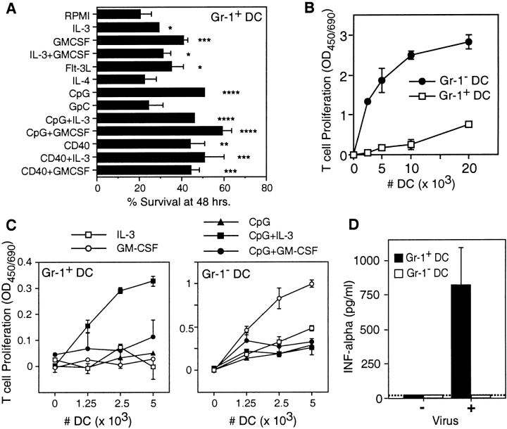
Human plasmacytoid dendritic cells (pDCs) are major producers of IFNalpha, are activated by CpG motifs, and are believed to enter lymph nodes (LNs) via L-selectin dependent extravasation across high endothelial venules. To identify a similar murine DC type, CD11c(+) cells in the LNs of L-selectin-deficient and control BALB/c mice were compared, revealing a population of CD11c(+)CD11b(-) cells that is reduced 85% in the LNs of L-selectin-deficient mice. These cells are Gr-1(+)B220(+)CD19(-), either CD4(+) or CD8(+), and localize within T cell zones of LNs. Freshly isolated CD11c(+)Gr-1(+) cells express major histocompatibility complex class II at low levels, display a plasmacytoid morphology, and survive poorly in culture. Their survival is increased and they develop a DC-like morphology in interleukin 3 and CpG. Like human pDCs, CD11c(+)Gr-1(+) cells stimulate T cell proliferation after activation with CpG and produce IFNalpha after stimulation with influenza virus. These cells also display a strain-specific variation in frequency, being fivefold increased in the LNs of BALB/c relative to C57BL/6 mice. These CD11c(+)CD11b(-)B220(+)Gr-1(+) cells appear to be the murine equivalent of human pDCs.


Figures

Figure 1
Figure 1
Flow cytometric analysis of DC populations in mouse lymph nodes and spleens. Low density cells, prepared from the indicated organs and mouse strains by enzymatic digestion and purification on Metrizimide gradients, were incubated with blocking reagents, the indicated fluorochrome-conjugated Abs, and 7-AAD before analysis. All panels are gated on forward and side scatter to exclude debris and on low autofluorescent, 7-AAD cells. Horizontal lines in the CD11c histograms indicate gates used for dot plots immediately below. Numbered squares in L indicate gates used for subsequent subset analyses and FACS® purification. All panels are representative of at least three separate analyses.
Figure 2
Figure 2
Surface marker expression on splenic CD11c+Gr-1+ cells. Low density 129/Sv spleen cells were stained with anti-CD11c, Gr-1, and the indicated FITC-conjugated mAbs. In each panel, staining of CD11c+Gr-1+ cells (gate 2 in Fig. 1 L) is shown in bold. Unless otherwise indicated, thin solid lines indicated staining profiles of CD11chiGr-1 cells (gate 3 in Fig. 1 L). Staining profiles of CD11c cells (gate 1 in Fig. 1 L) are indicate by the number “1.” Dotted lines represent staining of isotype controls. All panels are representative of at least three separate analyses.
Figure 3
Figure 3
Presence of low density B220+Gr-1+ cells in BALB/c, C57BL/6, and Rag /− mice. Low density cells were prepared from the indicated organs of mice as described in the legend to Fig. 1, stained with the indicated mAbs, and gated on low autofluorescent, 7-AAD cells. (A and B) Presence of B220+Gr-1+ cells in the spleens of BALB/c and C57BL/6 mice, respectively. (C and D) Frequency of B220+Gr-1+ cells in the LNs of BALB/c and BALB/c-Rag−/− mice. The intensity of Gr-1 staining in top and bottom panels are not directly comparable due to differences methodology.
Figure 4
Figure 4
Morphology and localization of CD11c+Gr-1+ cells. (A–E) Cytospins of FACS®-purified CD11c+Gr-1+ cells were Giemsa-stained and photographed at 100×. (A and B) Appearance of freshly isolated CD11c+Gr-1+ cells. (C and D) CD11c+Gr-1+ cells after overnight culture in IL-3 and GM-CSF. (E) CD11c+Gr-1+ cells after activation in IL-3 and CpG for 40 h. (F) Confocal microscopy of BALB/c LN frozen section stained with anti–B220-FITC (green) and anti–Gr-1-PE (red). Colocalization of B220 and Gr-1 signals appears as yellow. (G) Higher magnification demonstrates individual B220+Gr-1+ cells within T cell zone.
Figure 5
Figure 5
Survival and activities of CD11c+Gr-1+ cells. CD11c+Gr-1+ (Gr-1+ DC) and CD11chiGr-1 (Gr-1- DC) cells were purified by FACS® from the spleens of 129/Sv mice. (A) Survival of CD11c+Gr-1+ cells after 48 h of culture in the indicated agents. (B) Proliferation of naive allogenic T cells incubated with the indicated numbers of freshly isolated CD11c+Gr-1+ or CD11chiGr-1 cells. (C) Proliferation of naive allogenic T cells incubated with cytokine and CpG activated CD11c+Gr-1+ or CD11chiGr-1 cells. A–C are representative of three independent experiments, each preformed in triplicate (mean ± SD; *P < 0.05; **P < 0.01; ***P < 0.005; ****P < 0.001). (D) IFNα production by CD11c+Gr-1+ or CD11chiGr-1 cells incubated 24 h in the presence of absence of 1.3 HAU/ml influenza virus (mean ± SD, n = 2). Dashed line, detection limit of the assay.

References

    1. Banchereau J., Steinman R.M. Dendritic cells and the control of immunity. Nature. 1998;392:245–252. - PubMed
    1. Pulendran B., Banchereau J., Maraskovsky E., Maliszewski C. Modulating the immune response with dendritic cells and their growth factors. Trends Immunol. 2001;22:41–47. - PubMed
    1. Steinman R.M., Cohn Z.A. Identification of a novel cell type in peripheral lymphoid organs of mice. I. Morphology, quantitation, tissue distribution. J. Exp. Med. 1973;137:1142–1162. - PMC - PubMed
    1. Steinman R.M., Pack M., Inaba K. Dendritic cells in the T-cell areas of lymphoid organs. Immunol. Rev. 1997;156:25–37. - PubMed
    1. Vremec D., Zorbas M., Scollay R., Saunders D.J., Ardavin C.F., Wu L., Shortman K. The surface phenotype of dendritic cells purified from mouse thymus and spleeninvestigation of the CD8 expression by a subpopulation of dendritic cells. J. Exp. Med. 1992;176:47–58. - PMC - PubMed

Publication types

MeSH terms