. 2001 Nov;75(22):10643-50.
doi: 10.1128/JVI.75.22.10643-10650.2001.

An attenuating mutation in the 2A protease of swine vesicular disease virus, a picornavirus, regulates cap- and internal ribosome entry site-dependent protein synthesis

Affiliations

An attenuating mutation in the 2A protease of swine vesicular disease virus, a picornavirus, regulates cap- and internal ribosome entry site-dependent protein synthesis

Y Sakoda et al. J Virol. 2001 Nov.

Abstract

Virulent and avirulent strains of swine vesicular disease virus (SVDV), a picornavirus, have been characterized previously. The major determinants for attenuation have been mapped to specific residues in the 1D-2A-coding region. The properties of the 2A proteases from the virulent and avirulent strains of SVDV have now been examined. Both proteases efficiently cleaved the 1D/2A junction in vitro and in vivo. However, the 2A protease of the avirulent strain of SVDV was much less effective than the virulent-virus 2A protease at inducing cleavage of translation initiation factor eIF4GI within transfected cells. Hence the virulent-virus 2A protease is much more effective at inhibiting cap-dependent protein synthesis. Furthermore, the virulent-virus 2A protease strongly stimulated the internal ribosome entry sites (IRESs) from coxsackievirus B4 and from SVDV, while the avirulent-virus 2A protease was significantly less active in these assays. Thus, the different properties of the 2A proteases from the virulent and avirulent strains of SVDV in regulating protein synthesis initiation reflect the distinct pathogenic properties of the viruses from which they are derived. A single amino acid substitution, adjacent to His21 of the catalytic triad, is sufficient to confer the characteristics of the virulent-strain 2A protease on the avirulent-strain protease. It is concluded that the efficiency of picornavirus protein synthesis, controlled directly by the IRES or indirectly by the 2A protease, can determine virus virulence.


Figures

FIG. 1
FIG. 1
Structure of the SVDV cDNA plasmids used in this study. The region of the SVDV genome that contains the determinants of virulence is included within a Bst1107I-BssHII restriction enzyme cDNA fragment including the coding sequence for the whole of 1D and the N-terminal region of the 2A protease. Regions of the genome from the pathogenic J1 strain (hatched bars) and the attenuated 00 strain (open bars) were amplified by PCR and cloned as EcoRI (R)-BamHI (B) fragments into the vector pGEM3Z under the control of a T7 promoter (●). The SVDV IRES (solid bar) was within a KpnI (K)-MscI (M) fragment. The c-myc epitope tag (checkered bars) was attached to the N terminus of 1D. The amino acid differences (residues 20 and 126) within the 2A protease between the J1 and 00 strains of SVDV are shown. The 1D coding sequence of the 00 strain plasmid had two amino acid substitutions compared to the published sequence (C to Y, residue 69, and Y to C, residue 219) while the J1 strain 1D sequence had a single amino acid substitution (K to E, residue 266). These mutations were different for different isolates of the same plasmids (A to C) and hence were probably generated in the PCR.
FIG. 2
FIG. 2
In vitro analysis of SVDV 1D-2A expression and self-cleavage activity. The indicated plasmids were used to program in vitro TNT reactions. Samples from the reactions, taken at 30 and 120 min as indicated, were analyzed by SDS-PAGE and autoradiography. Uncleaved precursor 1D-2A and mature products 1D and 2A are identified, as is the uncleaved truncated product 1DΔ-2AΔ, generated by the deletion mutant lacking the 1D/2A cleavage site (Fig. 1).
FIG. 3
FIG. 3
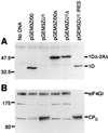
The SVDV 2A protease from strain J1, but not that from strain 00, induces efficient eIF4GI cleavage and inhibits its own synthesis in BHK cells. The indicated plasmids were transfected into vTF7-3-infected BHK cells. After 20 h, cell lysates were prepared and analyzed by SDS-PAGE and immunoblotting using antibodies specific for the c-myc epitope tag on the N terminus of 1D (A) and for the C terminus of eIF4GI (B). CPC, C-terminal cleavage product of eIF4GI. Note that the product of about 175 kDa that reacts with the anti-eIF4GI antisera is characteristic of BHK cell extracts (see also reference 31); it is lost following eIF4GI cleavage and may be a breakdown product of eIF4GI.
FIG. 4
FIG. 4
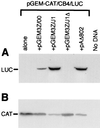
Differential inhibition of cap-dependent protein synthesis and IRES activation by the SVDV 2A proteases. The indicated test plasmids (Fig. 1) were cotransfected with reporter plasmid pGEM-CAT/CB4/LUC into vTF7-3-infected BHK cells. After 20 h cell extracts were prepared and analyzed by SDS-PAGE and immunoblotting using antibodies specific for LUC (A) and CAT (B). Plasmid pAΔ802 expresses the PV 2A protease.
FIG. 5
FIG. 5
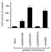
Quantitation of CB4 IRES activation by enterovirus 2A proteases in BHK cells. Reporter plasmid pGEM-CAT/CB4/LUC was transfected alone or with the indicated plasmids into vTF7-3-infected BHK cells. LUC activity within cell extracts was determined using a luciferase assay kit (Promega) and luminometer. Results shown are the means ± standard deviations from up to five separate determinations. In each experiment, the LUC expression obtained from the reporter plasmid alone was set at 100% and the relative activities obtained in the presence of the test plasmids were calculated. Extracts were also analyzed by SDS-PAGE and immunoblotting for CAT and LUC as for Fig. 4, and the results were mutually consistent in each case (data not shown).
FIG. 6
FIG. 6
Differential activation of the SVDV IRES by SVDV 2A proteases. (A) Structures of reporter plasmids pGEM-CAT/SVDJ(+)/LUC (pC/SVDJ(+)/L), pGEM-CAT/SVDJ(−)/LUC (pC/SVDJ(−)/L), and pGEM-CAT/SVD00(+)/LUC (pC/SVD00(+)/L). The orientation of the SVDV IRES and the source of SVDV cDNA (00, open box; J1, solid box) are indicated. (B) The reporter plasmids were transfected alone or with the indicated plasmids into vTF7-3-infected BHK cells. LUC activity was determined within cell extracts using a LUC assay kit (Promega) and luminometer. Results shown are the means ± standard deviations from three separate determinations. In each experiment, the LUC expression obtained from the reporter plasmid alone was set at 100% and the relative activities obtained in the presence of the test plasmids were calculated. The expression of LUC from pC/SVDJ(−)/L alone was about 1% of that obtained from pC/SVDJ(+)/L alone.
FIG. 7
FIG. 7
Residue 20 within the SVDV 2A protease determines its activity in BHK cells. (A) The indicated plasmids were transfected into vTF7-3-infected BHK cells as for Fig. 3. Cell extracts were analyzed for the integrity of eIF4GI by SDS-PAGE and immunoblotting. The full-length protein and its C-terminal cleavage product are indicated. (B) The dicistronic reporter plasmid containing the SVDV J1 IRES was transfected alone or with the indicated test plasmids into BHK cells (as for Fig. 6). Samples of cell extract were analyzed by SDS-PAGE and immunoblotting with anti-LUC antisera. The extracts were also analyzed for LUC expression by LUC assay. (C) From three separate experiments, performed as described for panel B, the level of LUC expression was measured by LUC assay with a luminometer. The activity observed from the reporter plasmid alone was set at 100% in each case, and other data were compared to it. The results presented are means ± standard deviations.

References

    1. Bazan J R, Fletterick R J. Viral cysteine proteases are homologous to the trypsin-like family of serine proteases: structural and functional implications. Proc Natl Acad Sci USA. 1988;85:7872–7876. - PMC - PubMed
    1. Belsham G J, Jackson R J. Translation initiation on picornavirus RNA. In: Sonenberg N, Hershey J W B, Mathews M B, editors. Translational control of gene expression. Monograph 39. Cold Spring Harbor, N.Y: Cold Spring Harbor Laboratory Press; 2000. pp. 869–900.
    1. Belsham G J, McInerney G M, Ross-Smith N. Foot-and-mouth disease virus 3C protease induces cleavage of translation initiation factors eIF4A and eIF4G within infected cells. J Virol. 2000;74:272–280. - PMC - PubMed
    1. Bernstein H D, Sonenberg N, Baltimore D. Poliovirus mutant that does not selectively inhibit host cell protein synthesis. Mol Cell Biol. 1985;5:2913–2923. - PMC - PubMed
    1. Borman A M, Le Mercier P, Girard M, Kean K M. Comparison of picornaviral IRES-driven internal initiation in cultured cells of different origins. Nucleic Acids Res. 1997;25:925–932. - PMC - PubMed

Publication types

LinkOut - more resources