. 2001 Nov;21(22):7641-52.
doi: 10.1128/MCB.21.22.7641-7652.2001.

Mechanisms of CAS substrate domain tyrosine phosphorylation by FAK and Src

Affiliations

Mechanisms of CAS substrate domain tyrosine phosphorylation by FAK and Src

P J Ruest et al. Mol Cell Biol. 2001 Nov.

Abstract

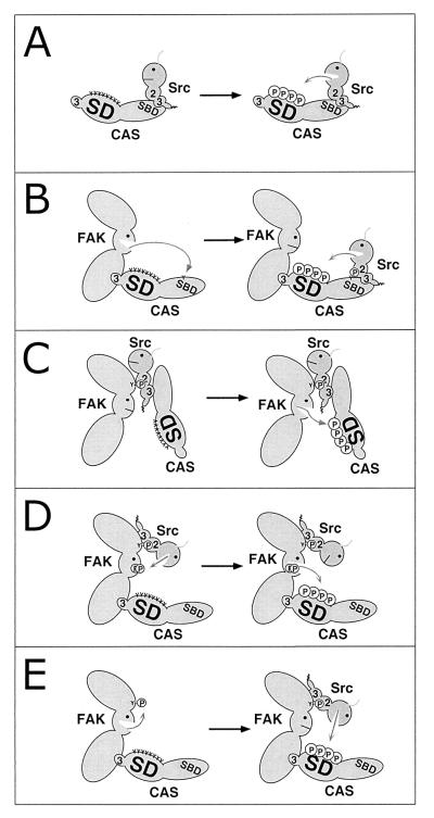
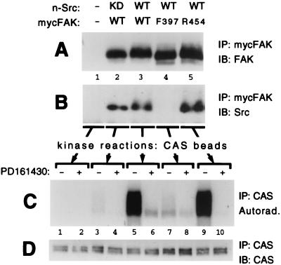
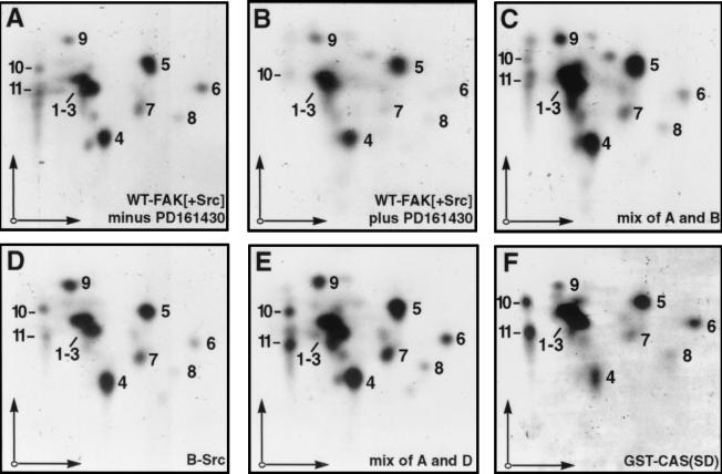
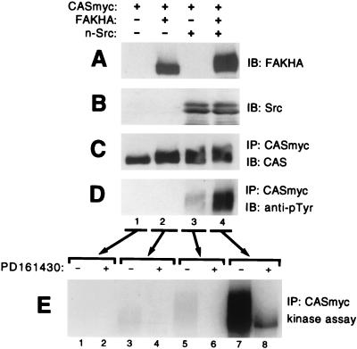
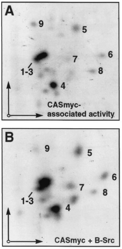
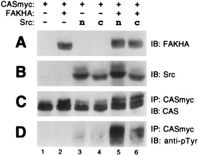
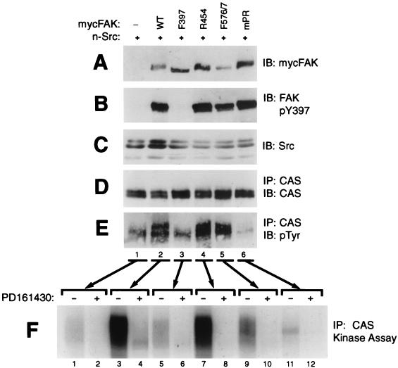
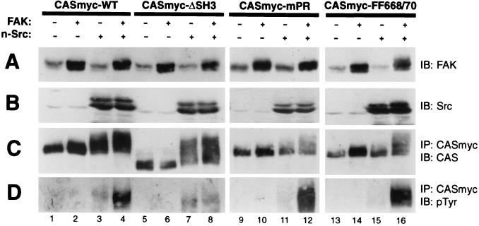
Tyrosine phosphorylation of CAS (Crk-associated substrate, p130(Cas)) has been implicated as a key signaling step in integrin control of normal cellular behaviors, including motility, proliferation, and survival. Aberrant CAS tyrosine phosphorylation may contribute to cell transformation by certain oncoproteins, including v-Crk and v-Src, and to tumor growth and metastasis. The CAS substrate domain (SD) contains 15 Tyr-X-X-Pro motifs, which are thought to represent the major tyrosine phosphorylation sites and to function by recruiting downstream signaling effectors, including c-Crk and Nck. CAS makes multiple interactions, direct and indirect, with the tyrosine kinases Src and focal adhesion kinase (FAK), and as a result of this complexity, several plausible models have been proposed for the mechanism of CAS-SD phosphorylation. The objective of this study was to provide experimental tests of these models in order to determine the most likely mechanism(s) of CAS-SD tyrosine phosphorylation by FAK and Src. In vitro kinase assays indicated that FAK has a very poor capacity to phosphorylate CAS-SD, relative to Src. However, FAK expression along with Src was found to be important for achieving high levels of CAS tyrosine phosphorylation in COS-7 cells, as well as recovery of CAS-associated Src activity toward the SD. Structure-functional studies for both FAK and CAS further indicated that FAK plays a major role in regulating CAS-SD phosphorylation by acting as a docking or scaffolding protein to recruit Src to phosphorylate CAS, while a secondary FAK-independent mechanism involves Src directly bound to the CAS Src-binding domain (SBD). Our results do not support models in which FAK either phosphorylates CAS-SD directly or phosphorylates CAS-SBD to promote Src binding to this site.


Figures

FIG. 1
FIG. 1
Models for CAS substrate domain phosphorylation by FAK or Src (see text for details). SD, substrate domain; SBD, Src-binding domain; 2, SH2 domain; 3, SH3 domain; Y, tyrosine; encircled P, phosphorylated tyrosine; visage, kinase domain.
FIG. 2
FIG. 2
CAS phosphorylation by FAK immunoprecipitates is largely due to coprecipitating Src. Myc-tagged FAK variants (either WT, F397, or R454) were expressed in COS-7 cells along with either WT n-Src or a kinase-dead (KD) n-Src mutant, as indicated. Control cells were transfected with empty expression plasmid (−). Cell lysates were incubated with anti-Myc antibody 9E10, and immunoprecipitates (IP) were formed with protein A-Sepharose beads that had been preadsorbed with mouse CAS protein which served as substrate for kinase reactions. After incubation with the lysates, the beads were split three ways and utilized for immunoblot (IB) assessment of FAK recovery (A) or coprecipitating Src recovery (B) or for kinase assays (C). The kinase reactions, utilizing [γ-32P]ATP, were carried out in the presence or absence of 44 μM PD161430, a Src-selective inhibitor. Reactions were stopped by boiling in SDS buffer, and CAS was reimmunoprecipitated to eliminate FAK, which comigrates with CAS on gels. After SDS-PAGE, CAS phosphorylation was visualized by autoradiography and quantitated by phosphorimage analysis. (D) The reimmunoprecipitated CAS was also subjected to immunoblot analysis to show near-equal recovery.
FIG. 3
FIG. 3
Tryptic phosphopeptide maps of mouse CAS phosphorylated in vitro. 32P-labeled CAS proteins phosphorylated in vitro by various kinase preparations were separated by SDS-PAGE, recovered from gels, and subjected to two-dimensional tryptic phosphopeptide mapping analysis. Electrophoretic separation (horizontal arrows) was in pH 1.9 buffer and chromatographic separation (vertical arrows) was in phosphochromo buffer. (A and B) CAS phosphorylated by WT-FAK immunoprecipitates from COS-7 cell lysates where n-Src was coexpressed in the absence (A) or presence (B) of the PD161439 Src inhibitor (i.e., lanes 5 and 6 in Fig. 2C). (C) Mix of samples A and B. (D) CAS phosphorylated by recombinant baculovirus-expressed c-Src (B-Src). (E) Mix of samples A and D. (F) GST-CAS(SD) phosphorylated by B-Src. For each map, ∼500 Cerenkov cpm were loaded on the thin-layer cellulose plates, and autoradiographic exposure was for 5 to 7 days.
FIG. 4
FIG. 4
FAK and n-Src cooperate to promote cellular CAS tyrosine phosphorylation and enhance CAS-associated kinase activity. Myc-tagged CAS (CASmyc) was expressed in COS-7 cells either alone, with HA-tagged FAK (FAKHA), with n-Src, or with both FAKHA and n-Src, and mycCAS phosphotyrosine content and associated kinase activity were determined. Equal total protein amounts of the cell lysates were used for immunoblot (IB) assessment of either FAKHA (A) or Src (B) expression using anti-HA antibody 12CA5 or anti-Src antibody 327, respectively. The remaining lysates (equal protein amounts) were used to immunoprecipitate (IP) CASmyc by using anti-Myc antibody 9E10 for the CAS phosphorylation analyses. (C) Near-equal CASmyc recovery was indicated by immunoblotting the immunoprecipitates with CAS-TL antibody. (D) Cellular phosphotyrosine levels of CASmyc were determined by immunoblotting equal portions of the CASmyc immunoprecipitates with antiphosphotyrosine antibody 4G10. (E) Kinase reactions were carried out in the presence of [γ-32P]ATP and either the presence (+) or absence (−) of 44 μM PD161430 on another equal portion of the mycCAS immunoprecipitates. Reactions were stopped by boiling in SDS buffer and CASmyc was reimmunoprecipitated to eliminate comigrating FAK. After SDS-PAGE, CAS phosphorylation was visualized by autoradiography.
FIG. 5
FIG. 5
Tryptic phosphopeptide maps of CASmyc phosphorylated by coprecipitating kinases. (A) WT-CASmyc coexpressed in COS-7 cells with FAK and n-Src and phosphorylated by coprecipitating kinases (i.e., lane 7 in Fig. 4E). (B) CASmyc expressed alone in COS-7 cells and phosphorylated by baculovirus-expressed Src (B-Src). For each map, ∼250 Cerenkov cpm were loaded on the thin-layer cellulose plates and autoradiographic exposure was for 7 days. Electrophoretic and chromatographic separation conditions were the same as for the maps shown in Fig. 3.
FIG. 6
FIG. 6
c-Src behaves similarly to n-Src in cooperating with FAK to promote CAS tyrosine phosphorylation. c-Src, which lacks the six-amino-acid nSrc insert in the SH3 domain, was compared to n-Src in its ability to promote CASmyc tyrosine phosphorylation in transfected COS-7 cells both in the presence or absence of FAKHA. See Fig. 4A to D legend for details. IB, immunoblot; IP, immunoprecipitate.
FIG. 7
FIG. 7
FAK structure/function requirements for FAK/Src-promoted CAS tyrosine phosphorylation. n-Src was expressed in COS-7 cells either alone or with a Myc-tagged FAK variant (either WT, F397, R454, F576/F577, or mPR [A712/A715/A873/A876]) and endogenous CAS phosphotyrosine content and associated kinase activity were assessed. Equal total protein amounts of the cell lysates were used for immunoblot (IB) assessment of either mycFAK expression using anti-Myc antibody 9E10 (A), FAK Tyr-397 phosphorylation using phosphospecific antibody pFAK397 (B), or Src expression using anti-Src antibody 327 (C). The remaining lysates (equal protein amounts) were used to immunoprecipitate (IP) endogenous CAS using CAS-TL antibody for the CAS phosphorylation analyses. (D) Near-equal CAS recovery was indicated by immunoblotting the immunoprecipitates with CAS-TL antibody. (E) Cellular phosphotyrosine levels of CAS were determined by immunoblotting equal portions of the CAS immunoprecipitates with anti-phosphotyrosine antibody 4G10. (F) Kinase reactions were carried out in the presence of [γ-32P]ATP and either the presence (+) or absence (−) of 44 μM PD161430 on another equal portion of the CAS immunoprecipitates. Reactions were stopped by boiling in SDS buffer, and CAS was reimmunoprecipitated to eliminate comigrating FAK. After SDS-PAGE, CAS phosphorylation was visualized by autoradiography.
FIG. 8
FIG. 8
CAS structure/function requirements for FAK/Src-promoted CAS tyrosine phosphorylation. Myc-tagged CAS (CASmyc) variants (either WT, ΔSH3 [deletion of SH3 domain], mPR [RAAASPP mutation of RPLPSPP motif], or F668/F670) were expressed in COS-7 cells either alone, with FAK (untagged), with n-Src, or with both FAK and n-Src, and the resulting phosphotyrosine content of the CASmyc variants was assessed. Equal total protein amounts of the cell lysates were used for immunoblot (IB) assessment of either FAK (A) or Src (B) expression using anti-FAK C20 antibody or anti-Src antibody 327, respectively. The remaining lysates (equal protein amounts) were used to immunoprecipitate (IP) CASmyc using anti-Myc antibody 9E10 for the CAS phosphotyrosine analyses. (C) Near-equal CASmyc recovery was indicated by immunoblotting the immunoprecipitates with CAS-B antibody. (D) Cellular phosphotyrosine levels of CASmyc were determined by immunoblotting equal portions of the immunoprecipitates with anti-phosphotyrosine antibody 4G10.

References

    1. Alexandropoulos K, Baltimore D. Coordinate activation of c-Src by SH3- and SH2-binding sites on a novel p130Cas-related protein, Sin. Genes Dev. 1996;10:1341–1355. - PubMed
    1. Almeida E A, Ilic D, Han Q, Hauck C R, Jin F, Kawakatsu H, Schlaepfer D D, Damsky C H. Matrix survival signaling: from fibronectin via focal adhesion kinase to c-Jun NH2-terminal kinase. J Cell Biol. 2000;149:741–754. - PMC - PubMed
    1. Astier A, Avraham H, Manie S N, Groopman J, Canty T, Avraham S, Freedman A S. The related adhesion focal tyrosine kinase is tyrosine-phosphorylated after β1 integrin stimulation in B cells and binds to p130cas. J Biol Chem. 1997;272:228–232. - PubMed
    1. Astier A, Manie S N, Avraham H, Hirai H, Law S F, Zhang Y, Golemis E A, Fu Y, Druker B J, Haghayeghi N, Freedman A S, Avraham S. The related adhesion focal tyrosine kinase differentially phosphorylates p130Cas and the Cas-like protein, p105HEF1. J Biol Chem. 1997;272:19719–19724. - PubMed
    1. Auvinen M, Paasinen-Sohns A, Hirai H, Andersson L C, Hölttä E. Ornithine decarboxylase- and ras-induced cell transformations: reversal by protein tyrosine kinase inhibitors and role of pp130Cas. Mol Cell Biol. 1995;15:6513–6525. - PMC - PubMed

Publication types