. 2001 Nov;21(22):7747-60.
doi: 10.1128/MCB.21.22.7747-7760.2001.

RasGAP-associated endoribonuclease G3Bp: selective RNA degradation and phosphorylation-dependent localization

Affiliations

RasGAP-associated endoribonuclease G3Bp: selective RNA degradation and phosphorylation-dependent localization

H Tourrière et al. Mol Cell Biol. 2001 Nov.

Abstract

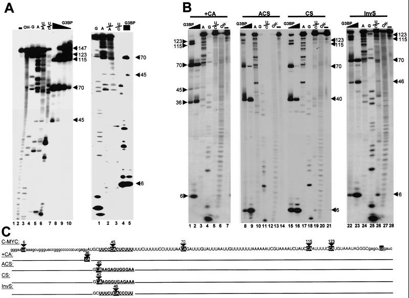
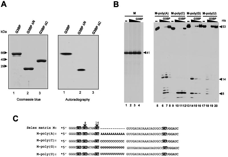
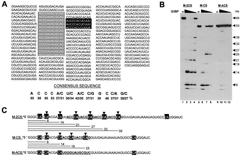
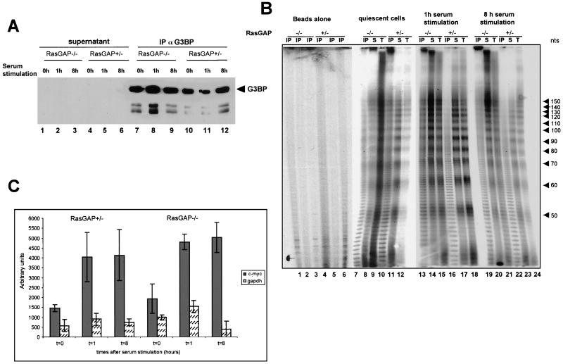
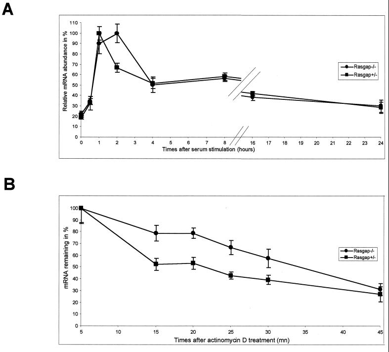
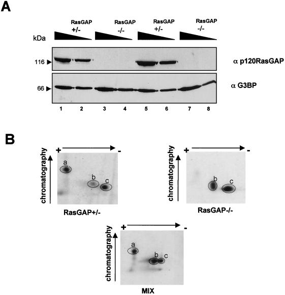
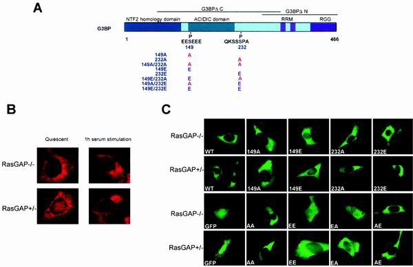
Mitogen activation of mRNA decay pathways likely involves specific endoribonucleases, such as G3BP, a phosphorylation-dependent endoribonuclease that associates with RasGAP in dividing but not quiescent cells. G3BP exclusively cleaves between cytosine and adenine (CA) after a specific interaction with RNA through the carboxyl-terminal RRM-type RNA binding motif. Accordingly, G3BP is tightly associated with a subset of poly(A)(+) mRNAs containing its high-affinity binding sequence, such as the c-myc mRNA in mouse embryonic fibroblasts. Interestingly, c-myc mRNA decay is delayed in RasGAP-deficient fibroblasts, which contain a defective isoform of G3BP that is not phosphorylated at serine 149. A G3BP mutant in which this serine is changed to alanine remains exclusively cytoplasmic, whereas a glutamate for serine substitution that mimics the charge of a phosphorylated serine is translocated to the nucleus. Thus, a growth factor-induced change in mRNA decay may be modulated by the nuclear localization of a site-specific endoribonuclease such as G3BP.


Figures

FIG. 1
FIG. 1
G3BP specifically cleaves wt and c-myc 3′ UTR mutants at CA sites. (A) 5′ 32P-labeled c-myc 3′ UTR transcripts (left panel, lane 1) were digested, using the RNA sequencing kit, with alkaline hydrolysis (lane 2), RNase T1 (cleaves 3′ to G residues) (lane 3), RNase U2 (cleaves 3′ to A residues) (lane 4), RNase Phy M (hydrolyses phosphodiester bonds 3′-adjacent to A and U residues [U/A]) (lane 5), RNase B. cereus (cleaves 3′ to pyrimidines U/C) (lane 6), or purified recombinant G3BP at concentrations of 8 pmol (lane 7), 4 pmol (lane 8), 2.4 pmol (lane 9), or 0.8 pmol (lane 10). Equivalent samples in lanes 3 to 7 were subjected to a short run electrophoresis (right panel, lanes 1 to 5, respectively). (B) 5′ 32P-labeled +CA (lanes 1 to 7), ACS (lanes 8 to 14), CS (lanes 15 to 21) and InvS (lanes 22 to 28) c-myc 3′ UTR mutants were either left unreacted (lanes 7, 14, 21, and 28) or digested with 2.4 pmol (lanes 1, 8, 15 and 22) or 8 pmol (lanes 2, 9, 16, and 23) of recombinant G3BP, RNase U2 (lanes 3, 10, 17, and 24), RNase T1 (lane 4, 11, 18, and 25), RNase B. cereus (lane 5, 12, 19, and 26), or alkaline hydrolysis (6, 13, 20, and 27). (C) Sequence of the c-myc 3′ UTR. Sequences from position 40 to 52 relative to the 5′ end which were mutated in ACS, CS, and InvS transcripts are noted in bold characters and underlined. The cleavage sites at CA dinucleotides are indicated by arrows. Nucleotides shown in lowercase characters are present in the transcription vector.
FIG. 2
FIG. 2
G3BP RNA binding activity and targeted cleavage by homopolymers. (A) Coomassie blue staining (left panel) of recombinant wt G3BP (lane 1), NH2-terminally truncated G3BP (lane 2), and COOH-terminally truncated G3BP (lane 3) purified after overexpression using a baculovirus system (see Materials and Methods). Right panel, autoradiography of Northwestern analysis of the same proteins, using 32P-labeled c-myc 3′ UTR as a probe. (B) 5′-end-labeled transcripts harboring constant regions either alone (M, lanes 1 to 4), or separated with 12 A residues [M-poly(A), lanes 5 to 8], 12 C residues [M-poly(C), lanes 9 to 12], 12 G residues [M-poly(G), lanes 13 to 16], or 12 U residues [M-poly(U), lanes 17 to 20] were digested with 8 pmol (lanes 2, 6, 10, 14, and 18), 2.4 pmol (lanes 3, 7, 11, 15, and 19), or 0.8 pmol (lanes 4, 8, 12, 16, and 20) of purified recombinant G3BP. (C) Sequences of the various transcripts. G3BP cleavage sites at CA dinucleotides are indicated by arrows. Homopolymers between constant regions are in boldface, and shading refers to empty region.
FIG. 3
FIG. 3
In vitro selection and amplification of high-affinity RNA target sequences for G3BP. (A) The sequences of individual clones after five cycles are shown. A consensus sequence was derived from the score of each nucleotide in the randomized sequence. Sequences that match the deduced consensus or deviate by 1 or 2 nt are boxed in grey. Sequences that are present in the c-myc 3′ UTR are boxed in black. (B) 5′-end-labeled transcripts containing constant regions separated by two copies (M-2CS, lanes 1 to 4), or one copy (M-CS, lanes 5 to 8) of the consensus sequence or its complementary sequence (M-ACS, lanes 9 to 12) were digested with 8 pmol (lanes 2, 6, and 10), 2.4 pmol (lanes 3, 7, and 11), or 0.8 pmol (lanes 4, 8, and 12) of purified recombinant G3BP. (C) Sequences of the various transcripts. The consensus sequence and its complementary sequence are noted in bold characters and underlined. G3BP cleavage sites at CA dinucleotides are indicated by arrows.
FIG. 4
FIG. 4
Cellular RNAs associated with G3BP. (A) G3BP was affinity purified by an anti-G3BP antibody immobilized on protein G-Sepharose, from total extracts of quiescent RasGAP−/− (lane 7) and RasGAP+/− (lane 10) cells or following serum stimulation for 1 h (lanes 8 and 11, respectively) or 8 h (lanes 9 and 12, respectively). Immunoprecipitates and corresponding supernatants (lanes 1 to 6) were analyzed by immunoblotting using anti-G3BP antibodies. (B) The poly(A) tails of RNAs from immunoprecipitates (IP) (lanes 7, 10, 13, 16, 19, and 22), supernatant (S) (lanes 8, 11, 14, 17, 20, and 23), and total extracts (T) (lanes 9, 12, 15, 18, 21, and 24) of quiescent RasGAP−/− (lanes 7 to 9) and RasGAP+/− (lanes 10 to 12) cells or those which were serum stimulated for 1 h (lanes 13 to 15 and lanes 16 to 18, respectively) or 8 h (lanes 19 to 21 and 22 to 24, respectively) were determined as described in Materials and Methods. (C) RNAs associated with affinity-purified G3BP from RasGAP−/− and RasGAP+/− quiescent cells or those cells which were serum stimulated for 1 and 8 h were extracted. The c-myc (grey bars) and GAPDH (white dotted bars) mRNAs from immunoprecipitates were detected by RT-PCR using specific primers. The amplified products were analyzed on an agarose gel, visualized by ethidium bromide, and quantified by fluorography. Background RT-PCR amplification from immunoprecipitates with beads alone in the absence of anti-G3BP antibodies was subtracted from the plotted values. Error bars resulting from two independently performed experiments, each measured in triplicate, are shown.
FIG. 5
FIG. 5
(A) c-myc mRNA expression in RasGAP+/ −and RasGAP−/− cells. After 24 h of serum starvation, expression was stimulated by serum addition for the indicated number of hours. RNA isolated from cells harvested at each time point was reverse transcribed and assayed for c-myc and GAPDH cDNAs using real-time quantitative PCR. All values were scaled to the level obtained from GAPDH, considered as a constitutively expressed mRNA. One hundred percent RNA is arbitrarily assigned to the time point which gave the highest signal. (B) Comparison of c-myc mRNA decay in RasGAP+/− and RasGAP−/− cells. Quiescent cells were stimulated with serum for 1 h and treated with 5 μg of actinomycin D/ml. At various times thereafter, cells were harvested and total RNA was prepared. The level of c-myc mRNA was quantified as in panel A and plotted. Error bars resulting from three independent experiments, each measured in duplicate, are shown.
FIG. 6
FIG. 6
(A) Proteins contained in total extracts derived from two cultures of exponentially growing RasGAP+/− (lanes 1, 2, 5, and 6) and RasGAP−/− (lanes 3, 4, 7, and 8) cells were analyzed by immunoblot using anti-G3BP and anti-p120 GAP antibodies. Amounts of loaded proteins were 15 μg (lanes 2, 4, 6, and 8) and 30 μg (lanes 1, 3, 5, and 7). (B) Phosphotryptic peptide mapping of immunopurified 32P-labeled G3BP from dividing RasGAP+/− (left panel) and RasGAP−/− (right panel) cells. The bottom panel represents the analysis of the mixture of the samples in both panels.
FIG. 7
FIG. 7
(A) Schematic representation of G3BP structural domains: NTF2 homology domain (in blue), acidic domain (in green), and RRM domain (in pink). The positions of the major phosphorylation sites are indicated. (B) Indirect immunofluorescent staining of quiescent and serum stimulated RasGAP−/− (top panels) or RasGAP+/− (bottom panels) cells with anti-G3BP antibodies. (C) Cellular localization of GFP fusion proteins in RasGAP+/− and RasGAP−/− cells. Direct fluorescence of GFP (GFP), GFP-G3BP (WT), GFP-S149A (149A), GFP-S149E (149E), GFP-S232A (232A), GFP-S232E (232E), and GFP-double mutants (EA, EE, AE, and AA) were performed 20 h after transfection. Expression of fusion proteins was confirmed by immunoblot analysis using an anti-GFP antibody (data not shown).

References

    1. Bashkirov V I, Scherthan H, Solinger J A, Buerstedde J M, Heyer W D. A mouse cytoplasmic exoribonuclease (mXRN1p) with preference for G4 tetraplex substrates. J Cell Biol. 1997;136:761–773. - PMC - PubMed
    1. Belasco J, Brawerman G. Control of messenger RNA stability. San Diego, Calif: Academic Press; 1993.
    1. Bernstein P, Peltz S W, Ross J. The poly(A)-poly(A)-binding protein complex is a major determinant of mRNA stability in vitro. Mol Cell Biol. 1989;9:659–670. - PMC - PubMed
    1. Bernstein P, Ross J. Poly(A), poly(A) binding protein and the regulation of mRNA stability. Trends Biochem Sci. 1989;14:373–377. - PubMed
    1. Bernstein P L, Herrick D J, Prokipcak R D, Ross J. Control of c-myc mRNA half-life in vitro by a protein capable of binding to a coding region stability determinant. Genes Dev. 1992;6:642–654. - PubMed

Publication types

MeSH terms