. 2001 Oct;134(4):753-62.
doi: 10.1038/sj.bjp.0704305.

Pravastatin suppresses the interleukin-8 production induced by thrombin in human aortic endothelial cells cultured with high glucose by inhibiting the p44/42 mitogen activated protein kinase

Affiliations

Pravastatin suppresses the interleukin-8 production induced by thrombin in human aortic endothelial cells cultured with high glucose by inhibiting the p44/42 mitogen activated protein kinase

M Takata et al. Br J Pharmacol. 2001 Oct.

Abstract

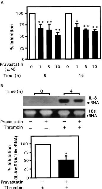
1. 3-Hydroxy-3-methylglutaryl co-enzyme A reductase inhibitors (statins) prevent the progression of atherosclerosis by lowering cholesterol. However, the effect of statins on the synthesis of pro-inflammatory cytokines from endothelial cells has not yet been fully investigated. Here, we examined the effect of pravastatin, one of the statins, on IL-8 synthesis induced by thrombin in human aortic endothelial cells (AoEC) cultured with high glucose concentrations. 2. Pravastatin significantly decreased the IL-8 synthesis induced by thrombin. 3. Pravastatin inhibited the p44/42 MAP kinase activity induced by thrombin, but did not inhibit the p38 MAP kinase activity. 4. Translocation of ras protein from the cytosol to plasma membrane was inhibited by pravastatin. 5. Pravastatin inhibit the activator protein-1 activity, but did not inhibit the activation of IkappaB-alpha. 6. Dominant negative ras inhibited the p44/42 MAP kinase activity induced by PMA. 7. Our results suggest that pravastatin inhibits IL-8 synthesis by blocking the ras-MAP (p44/42) kinase pathway rather than nuclear factor-kappaB. Pravastatin may prevent atherosclerosis not only by lowering cholesterol levels, but also by suppressing IL-8 synthesis in AoEC through the inhibition of p44/42 MAP kinase, and this may be more beneficial in diabetic patients than in non-diabetics.


Figures

Figure 1
Figure 1
Effect of thrombin on IL-8 synthesis and mRNA expression in human aortic endothelial cells (AoEC). (A) AoEC at passages 5 – 10 were cultured with medium containing 5.5 mM of glucose in 24-well collagen-coated plates. The cells were then stimulated with the indicated concentration of thrombin for 8 and 16 h. The concentration of IL-8 in the medium was measured by ELISA. (B) AoEC were cultured with medium containing 5.5 mM of glucose in 6 cm collagen-coated dishes. The cells were then serum-starved with medium containing 0.5% FCS for 16 h. After stimulation with thrombin (10 U ml−1) for 4 h, the expression of IL-8 mRNA was analysed by Northern blot analysis. The bands were analysed by NIH image. The data are representative of three different experiments (means±s.d.). *P<0.001 vs control (white bar).
Figure 2
Figure 2
Effect of pravastatin on the IL-8 production (A) and the IL-8 mRNA expression (B) induced by thrombin in human aortic endothelial cells (AoEC). (A) AoEC were cultured with media containing 5.5 mM of glucose in 24-well collagen-coated plates with various concentrations of pravastatin for 14 days. The cells were then stimulated with thrombin for 8 and 16 h. The concentration of IL-8 in the medium was measured by ELISA. (B) AoEC were cultured in 6 cm collagen-coated dishes with or without pravastatin (10 μM) for 14 days, and the cells were then serum-starved with medium containing 0.5% FCS for 16 h. After stimulation with thrombin (10 U ml−1) for 4 h, the expression of IL-8 mRNA was analysed by Northern blot analysis. The bands were analysed by NIH image, and the intensity of the IL-8 mRNA band was corrected with that of the 18s rRNA band. The data are representative of three different experiments (means±s.d.). *P<0.01 vs control (white bar), **P<0.001 vs control (white bar).
Figure 3
Figure 3
Thrombin activates the p44/42 MAP kinase and the p38 MAP kinase in human aortic endothelial cells (AoEC). AoEC were cultured with medium containing 5.5 mM glucose in 6 cm collagen-coated dishes before being serum-starved for 16 h with medium containing 0.5% FCS and then stimulated with thrombin (10 U ml−1) for 5, 10 and 15 min. Cell lysates were separated by 10% SDS – PAGE, transferred onto membranes, and blotted with phospho-specific p44/42 MAP kinase (Thr202 and Tyr204) antibody (A) or a phospho-p38 MAP kinase (Thr180 and Tyr182) antibody (B).
Figure 4
Figure 4
Effect of PD 98059 (A) or SB 203580 (B) on the IL-8 mRNA expression induced by thrombin in human aortic endothelial cells (AoEC). AoEC cultured with media containing 5.5 mM glucose were serum-starved with medium containing 0.5% FCS for 16 h. The cells were then treated for 1 h with PD 98059 (20 μM) (A), a specific inhibitor of MEK, or SB 203580 (20 μM) (B), an inhibitor of p38 MAP kinase. Next, they were stimulated with thrombin (10 U ml−1), and then the RNA was extracted. The expression of IL-8 mRNA was analysed by Northern blot analysis. The bands were analysed by NIH image, and the intensity of the IL-8 mRNA band was corrected with that of the 18s rRNA band. The data are representative of three different experiments (means±s.d.). *P<0.01 vs control (white bar), **P<0.05 vs control (white bar).
Figure 5
Figure 5
Effect of pravastatin on the activity of MAP kinases induced by thrombin in human aortic endothelial cells (AoEC). AoEC were cultured with or without pravastatin (10 μM) for 14 days before being serum-starved with medium containing 0.5% FCS for 16 h in 6 cm collagen-coated dishes and then stimulated with thrombin (10 U ml−1) for 15 min. Cell lysates were separated by 10% SDS – PAGE, transferred onto membranes, and blotted with phospho-specific p44/42 MAP kinase (Thr202 and Tyr204) antibody (A) or a phospho-p38 MAP kinase (Thr180 and Tyr182) antibody (B). The bands were analysed by NIH image, and the intensity of each phospho-MAP kinase band was corrected with that of the respective MAP kinase band. The data are representative of three different experiments (means±s.d.). *P<0.001 vs control (white bar).
Figure 6
Figure 6
Effect of mevalonate (A) and FPP (B) on the activity of p44/42 MAP kinase induced by thrombin in human aortic endothelial cells (AoEC). AoEC were cultured with or without pravastatin (10 μM) or with pravastatin (10 μM) combined with mevalonate (400 μM) (A) or FPP (5 μM) (B), serum-starved by the media containing 0.5% FCS for 16 h in 6 cm collagen-coated dishes and then stimulated with thrombin (10 U ml−1) for 5 min. Cell lysates were separated by 10% SDS – PAGE, transferred onto membranes, and blotted with phospho-specific p44/42 MAP kinase (Thr202 and Tyr204) antibody. The bands were analysed by NIH image, and the intensity of each phospho-MAP kinase band was corrected with that of the respective MAP kinase band. The data are representative of three different experiments (means±s.d.). *P<0.05.
Figure 7
Figure 7
Effect of pravastatin on the processing of ras protein in human aortic endothelial cells (AoEC). AoEC were cultured with medium containing 5.5 mM of glucose in the presence or absence of pravastatin (10 μM) for 14 days. The cells were lysed with lysis buffer, and the cell lysates were fractionated by ultra-centrifugation. Cytosolic (supernatant) and plasma membrane (pellet) fractions were separated by 12% SDS – PAGE, transferred onto membranes and blotted with an Ha-ras antibody.
Figure 8
Figure 8
Effect of dominant negative ras protein on p44/42 MAP kinase activity induced by thrombin in human aortic endothelial cells (AoEC). A dominant negative H-ras (S17N) was transfected to AoEC using Lipofectin. After transfection, the AoEC were cultured with medium containing 10% FCS for 48 h, serum-starved by the media containing 0.5% FCS for 16 h, and then stimulated with thrombin (10 U ml−1) for 5 min. Cell lysates were separated by 10% SDS – PAGE, transferred onto membranes and blotted with phospho-specific p44/42 MAP kinase (Thr202 and Tyr204) antibody and an Ha-ras antibody. The bands were analysed by NIH image, and the intensity of each phospho-MAP kinase band was corrected with that of the respective MAP kinase band. The data are the representative of three different experiments (mean±s.d.). *P<0.001 vs control (white bar).
Figure 9
Figure 9
Changes in the thrombin induced-phosphorylation of IκB-α in human aortic endothelial cells (AoEC) treated with pravastatin, Calphostin C, or PD 98059. AoEC cultured with medium containing 5.5 mM glucose were treated with pravastatin (10 μM) for 14 days, or treated by PD 98059 (20 μM) for 1 h, or treated with Calphostin C (100 nM) for 1 h. Cells were stimulated with thrombin (10 U ml−1) for 15 min, respectively. Cell lysates were separated by 10% SDS – PAGE, transferred to membranes and blotted with a phospho-IκB-α (Ser32) antibody. The data are representative of three different experiments.
Figure 10
Figure 10
Effect of pravastatin on the binding of specific DNA sequences to AP-1 transcription factor complexes induced by thrombin in human aortic endothelial cells (AoEC). AoEC were cultured with medium containing 5.5 mM glucose in the presence or absence of pravastatin (10 μM) for 14 days, serum-starved by the media containing 0.5% FCS for 16 h in 6 cm collagen-coated dishes, and then stimulated with thrombin (10 U ml−1) for 1 h. Nuclear extracts were isolated, and used for gel mobility shift assay. A double-stranded DNA fragments containing the sequence of the binding site for the transcription factor AP-1 was labelled with digoxigenin-11-ddUTP by terminal transferase using a DIG gel shift kit (Roche). Nuclear proteins were then incubated with the DIG-labelled DNA fragment. The reaction mixture was loaded onto a 5% polyacrylamide gel and transferred onto a membrane. The bands of DNA-nuclear protein complex were detected with an anti-digoxigenin antibody (Roche). The bands were analysed by NIH image. The data are representative of three different experiments (means±s.d.). *P<0.001, **P<0.01.
Figure 11
Figure 11
Effect of high glucose and Calphostin C on p44/42 MAP kinase activity in human aortic endothelial cells (AoEC). AoEC were cultured with medium containing 5.5, 24 or 42.5 mM glucose for 14 days, serum-starved with medium containing 0.5% FCS for 16 h, and then treated with Calphostin C (100 nM) for 1 h. Cell lysates were separated by 10% SDS – PAGE, transferred to membranes and blotted with phospho-specific p44/42 MAP kinase (Thr202 and Tyr204) antibody. The bands were analysed by NIH image, and the intensity of each phospho-MAP kinase band was corrected with that of the respective MAP kinase band. The data are representative of three different experiments (mean±s.d.). *P<0.05, **P<0.01.
Figure 12
Figure 12
Effect of pravastatin on p44/42 MAP kinase activity induced by high glucose in human aortic endothelial cells (AoEC). AoEC were cultured with medium containing 5.5, 10 or 24 mM glucose in the presence or absence of pravastatin (10 μM) for 14 days, serum-starved by the medium containing 0.5% FCS for 16 h, and then stimulated with thrombin (10 U ml−1) for 15 min. Cell lysates were separated by 10% SDS – PAGE, transferred onto membranes and blotted with phospho-specific p44/42 MAP kinase (Thr202 and Tyr204) antibody. The data are representative of three different experiments (mean±s.d.).
Figure 13
Figure 13
Effect of dominant negative ras protein on p44/42 MAP kinase activity induced by PMA in human aortic endothelial cells (AoEC). A dominant negative H-ras (S17N) was transfected into AoEC using Lipofectin. After transfection, the AoEC were cultured with medium containing 10% FCS for 48 h. The cells were then serum-starved with the media containing 0.5% FCS for 16 h before being stimulated with PMA (100 nM) for 15 min. Cell lysates were separated by 10% SDS – PAGE, transferred onto membranes and blotted with phospho-specific p44/42 MAP kinase (Thr202 and Tyr204) antibody and an Ha-ras antibody. The bands were analysed by NIH image, and the intensity of each phospho-MAP kinase band was corrected with that of the respective MAP kinase band. The data are the representative of three different experiments (mean±s.d.). *P<0.001 vs control (white bar).

References

    1. ANRATHER D., MILLAN M.T., PALMETSHOFER A., ROBSON S.C., GECZY C., RITCHIE A.J., BACH F.H., EWENSTEIN B.M. Thrombin activates nuclear factor-kappaB and potentiates endothelial cell activation by TNF. J. Immunol. 1997;159:5620–5628. - PubMed
    1. BOISVERT W.A., SANTIAGO R., CURTISS L.K., TERKELTAUB R.A. A Leukocyte Homologue of the IL-8 Receptor CXCR-2 Mediates the Accumulation of Macrophages in Atherosclerotic Lesions of LDL Receptor-deficient Mice. J. Clin. Invest. 1998;101:353–363. - PMC - PubMed
    1. BACON K.B., WESTWICK J., CAMP R.D. Potent and specific inhibition of IL-8-, IL-1 alpha- and IL-1 beta-induced in vitro human lymphocyte migration by calcium channel antagonists. Biochem. Biophys. Res. Commun. 1989;165:349–354. - PubMed
    1. CERIELLO A., GIACOMELLO R., COLATUTTO A., TABOGA C., GONANO F. Increased prothrombin fragment 1+2 in type I diabetic patients. Haemostasis. 1992;22:50–51. - PubMed
    1. ENDRES M., LAUFS U., HUANG Z., NAKAMURA T., HUANG P., MOSKOWITZ M.A., LIAO J.K. Stroke protection by 3-hydroxy-3-methylglutaryl (HMG)-CoA reductase inhibitors mediated by endothelial nitric oxide synthase. Proc. Natl. Acad. Sci. U.S.A. 1998;95:8880–8885. - PMC - PubMed

MeSH terms