. 2001 Nov;2(11):1018-23.
doi: 10.1093/embo-reports/kve223. Epub 2001 Oct 17.

The cyclin B1 gene is actively transcribed during mitosis in HeLa cells

Affiliations

The cyclin B1 gene is actively transcribed during mitosis in HeLa cells

S Sciortino et al. EMBO Rep. 2001 Nov.

Abstract

In mammalian cells, the expression level of the cyclin B1 gene plays a critical role in the progression through mitosis. Here we demonstrate that the transcriptional activity of the human cyclin B1 promoter, as well as the rate of gene transcription, is high during mitosis. Indeed, the cyclin B1 promoter maintains an open chromatin configuration at the mitotic stage. Consistent with this, we show that the cyclin B1 promoter is occupied and bound to NF-Y during mitosis in vivo. Our results provide the first example of RNA polymerase II-dependent transcription during mitosis in mammalian cells.


Figures

None
Fig. 1. Transcriptional activity of the exogenous cyclin B1 promoter is high in the G2/M phases of the cell cycle. (A) DNA distribution analysis of propidium iodide-stained asynchronous (Async.) and MSO HeLa cells. (B) Northern blot analysis of CAT mRNA levels in asynchronous (Async.) and MSO cells. HeLa cells stably transfected with the CAT reporter gene fused to the 332 bp cyclin B1 promoter were synchronized by a double thymidine block. The filter was hybridized with CAT, murine cyclin B1 and human GAPDH cDNAs.
None
Fig. 2. The cyclin B1 promoter is accessible to restriction endonucleases in vivo. Nuclei isolated from asynchronous (Async.) (20 µg of DNA) and permeabilized MSO cells were partially digested with 100 U of BsaAI (A), 100 U of Sau96I (B), 200 U of XbaI (C) or 100 U of SalI (D). Purified DNA was fully digested with NcoI and XbaI when BsaAI was used for the partial digestion, and with NcoI when XbaI was used for the partial digestion. The probe used for the hybridization is shown at the bottom of each panel. Arrows mark the digested and undigested bands. The schematic diagram at the bottom of each panel indicates the position of the digested bands in the cyclin B1 promoter. The percent digestion is shown below each lane.
None
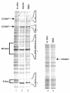
Fig. 3. The cyclin B1 promoter interacts with transcription factors during mitosis in vivo. Genomic footprinting of the non-coding strand of the human cyclin B1 promoter from asynchronous (Async.) and MSO HeLa cells (lanes 2 and 3), and of the coding strand of the human hsp70 promoter from MSO HeLa cells (lane 5). As a control, the DMS reactivity for DNA purified from asynchronous HeLa cells is shown (In vitro). The changes in DMS reactivity found in asynchronous and MSO cells are noted at the corresponding G residues by an asterisk (hypersensitivity) and by underlines (protection).
None
Fig. 4. NF-Y binds to the cyclin B1 promoter in vivo during mitosis. (A) DNA distribution analysis of propidium iodide-stained MSO and G1 HeLa cells. (B) Formaldehyde cross-linked chromatin was prepared from the same number of asynchronous (Async.), mitotic and G1 HeLa cells. Cross-linked chromatin from each sample was incubated with antibodies to NF-Y (lanes 2, 3, 8, 9, 13 and 14) and E2F1 (lanes 4, 10 and 15), or in the absence of antibody (none). Immunoprecipitates from each antibody were aliquoted and then analyzed by PCR with specific primers for the cyclin B1 or hsp70 promoters. A sample representing 0.02% of the total input chromatin (input) was included in the PCR analysis.
None
Fig. 5. Transcriptional activity of endogenous cyclin B1 promoter is high during mitosis. Asynchronous (Async.), mitotic and G1 phase HeLa cells were tested by a whole-cell run-on assay. Probes were cDNAs from the murine cyclin B1, A, E and D1 genes.

Similar articles

Cited by

References

    1. Baum B., Nishitani, H., Yanow, S. and Nurse, P. (1998) Cdc18 transcription and proteolysis couple S phase to passage through mitosis. EMBO J., 17, 5689–5698. - PMC - PubMed
    1. Bhattacharyya N., Dey, A., Minucci, S., Zimmer, A., John, S., Hager, G. and Ozato, K. (1997) Retinoid-induced chromatin structure alterations in the retinoic acid receptor β2 promoter. Mol. Cell. Biol., 17, 6481–6490. - PMC - PubMed
    1. Boyd K.E., Wells, J., Gutman, J., Bartley, S.M. and Farnham, P.J. (1998) c-Myc target gene specificity is determined by a post-DNA binding mechanism. Proc. Natl Acad. Sci. USA, 95, 13887–13892. - PMC - PubMed
    1. Clute P. and Pines, J. (1999) Temporal and spatial control of cyclin B1 destruction in metaphase. Nature Cell. Biol., 1, 82–87. - PubMed
    1. Cogswell J.P., Godlevski, M.M., Bonham, M., Bisi, J. and Babiss, L. (1995) Upstream stimulatory factor regulates expression of the cell cycle-dependent cyclin B1 gene promoter. Mol. Cell. Biol., 15, 2782–2790. - PMC - PubMed

Publication types