. 2001 Nov 1;21(21):8447-55.
doi: 10.1523/JNEUROSCI.21-21-08447.2001.

Microglial activation and dopaminergic cell injury: an in vitro model relevant to Parkinson's disease

Affiliations

Microglial activation and dopaminergic cell injury: an in vitro model relevant to Parkinson's disease

W Le et al. J Neurosci. .

Abstract

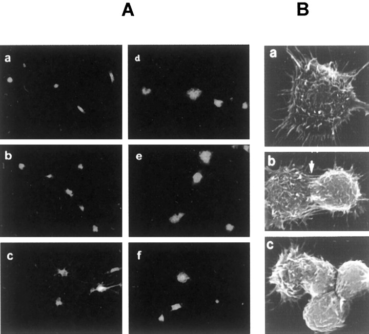
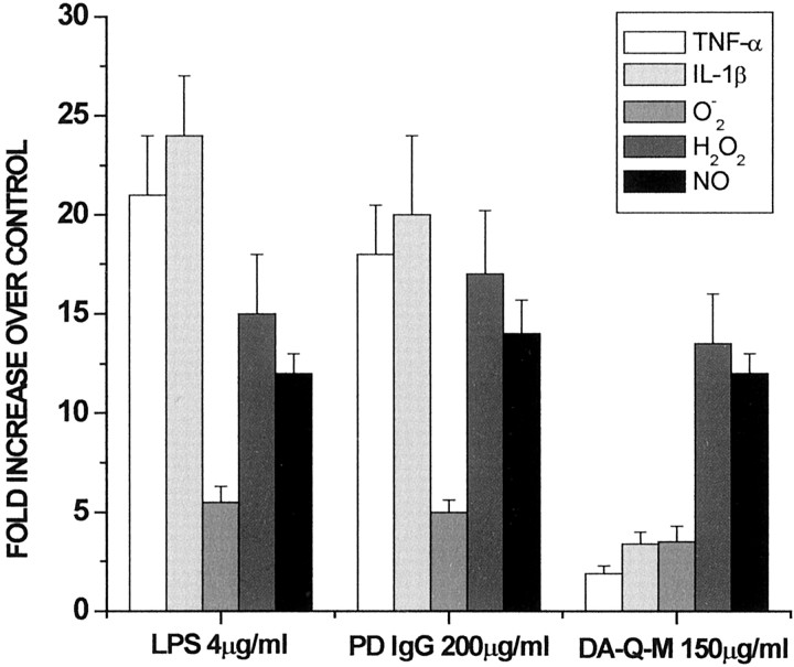
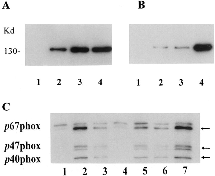
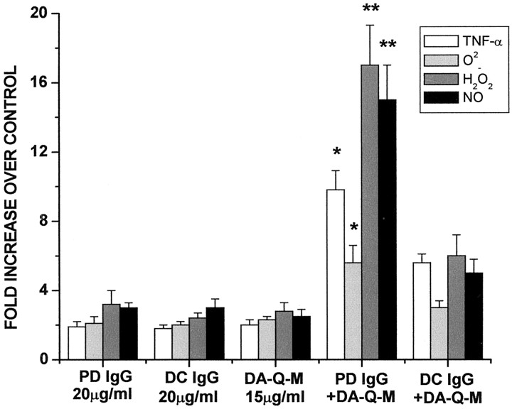
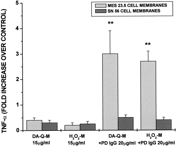
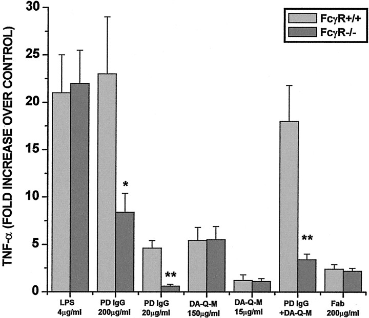
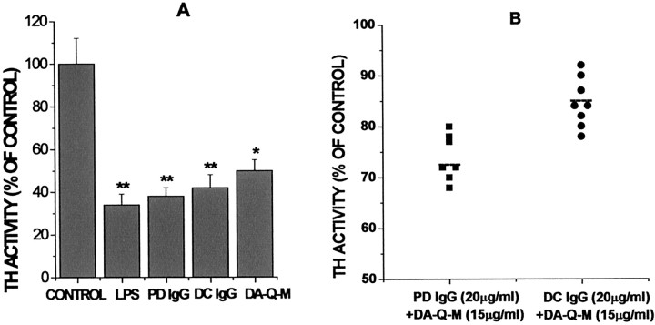
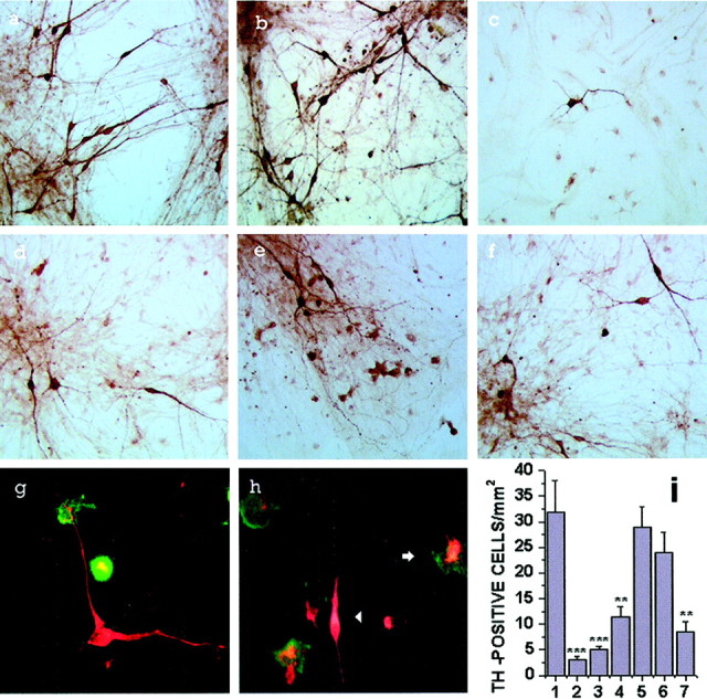
Microglial activation and oxidative stress are significant components of the pathology of Parkinson's disease (PD), but their exact contributions to disease pathogenesis are unclear. We have developed an in vitro model of nigral injury, in which lipopolysaccharide-induced microglial activation leads to injury of a dopaminergic cell line (MES 23.5 cells) and dopaminergic neurons in primary mesencephalic cell cultures. The microglia are also activated by PD IgGs in the presence of low-dose dopa-quinone- or H(2)O(2)-modified dopaminergic cell membranes but not cholinergic cell membranes. The activation requires the microglial FCgammaR receptor as demonstrated by the lack of activation with PD IgG Fab fragments or microglia from FCgammaR-/- mice. Although microglial activation results in the release of several cytokines and reactive oxygen species, only nitric oxide and H(2)O(2) appear to mediate the microglia-induced dopaminergic cell injury. These studies suggest a significant role for microglia in dopaminergic cell injury and provide a mechanism whereby immune/inflammatory reactions in PD could target oxidative injury relatively specifically to dopaminergic cells.


Figures

Fig. 1.
Fig. 1.
A, The morphology of rat microglial cells labeled with DIL-ac-LDL. Rat microglia were incubated for 2 d with vehicle (Aac), LPS (4 μg/ml) (Ad), high-dose PD IgG (200 μg/ml) (Ae), and high-dose DA-Q-M MES 23.5 cell membranes (150 μg/ml) (Af). Note that microglia after being treated with LPS, PD IgG, or DA-Q-M MES 23.5 cell membranes became larger and round. B, Scanning electron microscopy of microglia and MES 23.5 cells. Ba, Individual activated microglia showed spikes and cell-surface features. Bb,Bc, Activated microglia (left) in contact (arrow) with MES 23.5 cell (right).
Fig. 2.
Fig. 2.
Activating effects of LPS, PD IgG, and DA-Q-M membranes on microglia. Microglial activation was determined by measuring the levels of TNF-α, IL-1β, O2, H2O2, and NO in the culture media. Microglia were incubated with LPS (4 μg/ml), high-dose PD IgG (200 μg/ml; n = 7), and high-dose DA-Q-M membranes (150 μg/ml) for 2 d, and medium was collected for measurement of TNF-α and IL-1β levels. Some of the microglial cultures were stimulated with 1 μg/ml PMA for 2 hr before assaying the released levels of O2, H2O2, and NO.
Fig. 3.
Fig. 3.
iNOS (A, B) and NADPH oxidase (C) induction in activated microglia. A, B, iNOS was detected (A, B) by immunoblot with iNOS antibodies. A, Microglia were incubated with (1) vehicle, (2) high-dose (150 μg/ml) DA-Q-M MES 23.5 cell membranes, (3) high-dose (200 μg) PD IgG, and (4) LPS (4 μg/ml) for 2 d.B, Microglia were incubated with (1) vehicle, (2) low-dose (15 μg/ml) DA-Q-M membranes, (3) low-dose (20 μg/ml) PD IgG, and (4) low-dose DA-Q-M membranes plus IgG. Note that increased iNOS in microglia after treatment with DA-Q-M membranes, IgG and LPS, and a synergetic effect of DA-Q-M membranes + IgG on microglia iNOS.C, NADPH oxidase was detected by three antibodies (p67phox, p47phox, and p40phox) in microglia treated with (1) vehicle, (2) high-dose PD IgG, (3) high-dose DA-Q-M membranes, (4) trypsin-treated DA-Q-M membranes, (5) low-dose PD IgG, (6) low-dose DA-Q-M membranes, and (7) low-dose DA-Q-M membranes + PD IgG. Arrows indicate three isoforms of NADPH oxidase reacting with antibodies ofp67phox, p47phox, andp40phox, respectively.
Fig. 4.
Fig. 4.
Microglial activation by IgG from PD and disease control (DC). The levels of TNF-α, O2, H2O2, and NO in the microglial cultures treated with low-dose PD IgG (20 μg/ml), low-dose DC IgG (20 μg/ml), low-dose PD IgG + low-dose DA-Q-M MES 23.5 cell membranes (15 μg/ml) +, low-dose DC IgG + low dose DA-Q-M MES 23.5 cell membranes, and low-dose DA-Q-M MES 23.5 cell membranes alone. *p < 0.05 and **p < 0.01 compared with DC IgG +DA-Q-M.
Fig. 5.
Fig. 5.
The specificity of microglial activation induced by DA-Q-M- or H2O2-M membranes from MES 23.5 cells as compared with SN 56 cells. The cells were treated with DA-Q (50 μm) or H2O2 (10 μm) for 24 hr, and the cell membranes alone or in combination with PD IgG were incubated with rat microglia for 2 d. TNF-α levels in the culture medium were determined by ELISA. **p < 0.01 versus SN 56 cell membrane addition.
Fig. 6.
Fig. 6.
Role of FcR in microglial activation. Mouse microglia were purified from the brains of 4- to 5-d-old mice with intact FcR γ chain (FcγR+/+) or with deleted FcR γ chain (FcγR−/−). The microglia were incubated with LPS (4 μg/ml), high-dose PD IgG (200 μg/ml), low-dose PD IgG (20 μg/ml; n = 3), high-dose DA-Q-M membranes (150 μg/ml), low dose DA-Q-M membranes (15 μg/ml), low-dose PD IgG + dose DA-Q-M membranes, and high-dose Fab fragment of PD IgG (200 μg/ml; n = 3) for 2 d. Microglial activation was determined by TNF-α release in the culture medium. *p < 0.005 and **p< 0.001 versus control FcγR+/+ microglia.
Fig. 7.
Fig. 7.
Reactive microglia induced MES 23.5 cell injury. MES 23.5 cell injury was determined by measuring TH activity in the cocultures with microglia. A, Cocultures were treated with vehicle, LPS (4 μg/ml), high-dose PD IgG (200 μg/ml), high-dose DC IgG (200 μg/ml), and high-dose DA-Q-M MES 23.5 cell membranes (150 μg/ml). B, Specificity of low-dose PD IgG (20 μg/ml) + DA-Q-M membranes (15 μg/ml) induced MES 23.5 cell injury. Column 1, Low-dose PD IgG (20 μg/ml;n = 7) + low-dose DA-Q-M MES 23.5 cell membranes (15 μg/ml). Column 2, Low-dose DC IgG (n = 8) + low-dose DA-Q-M MES 23.5 cell membranes (15 μg/ml). Dashed lines represent the mean value of TH activity in the cocultures treated with PD IgG (n = 7) or DC IgG + DA-Q-M MES 23.5 cell membranes.p < 0.05; PD IgG + DA-Q-M membranes versus DC IgG + DA-Q-M membranes. *p < 0.01 and **p < 0.005 versus control cocultures.
Fig. 8.
Fig. 8.
Reactive microglia-induced primary mesencephalic cell injury. Representative photographs of TH immunostaining in primary mesencephalic cell cultures (a) or cocultures with microglia (bf) in addition of vehicle (b), LPS (4 μg/ml) (c), high-dose PD IgG (200 μg/ml) (d), high-dose DA-Q-M membranes (150 μg/ml) (e), and low-dose PD IgG (20 μg/ml) + low-dose DA-Q-M membranes (15 μg/ml) (f) for 2 d. Double staining of TH-positive neurons (red) and OX-42-positive microglia (green) in the vehicle-treated cocultures (g) and PD IgG-activated cocultures (h). Inh, note a reactive microglia surrounding and phagocytosing an injured dopaminergic neuron (arrow), and a TH-positive neuron with attenuated neurite and shortened cell body (arrowhead). I, Quantitative counting of TH-positive cells in cocultures. Control cocultures of primary mesencephalic cells with microglia treated with vehicle (column 1), 4 μg/ml LPS (column 2), high-dose PD IgG (200 μg/ml) (column 3), high-dose DA-Q-M membranes (150 μg/ml) (column 4), low-dose DA-Q-M membranes (15 μg/ml) (column 5), low-dose PD IgG (20 μg/ml) (column 6), and low-dose PD IgG + low-dose DA-Q-M membranes (column 7). *p < 0.05, **p < 0.01, and ***p < 0.001 versus control cocultures (column 1).

References

    1. Appel SH, Le WD, Tajti J, Haverkamp LJ, Engelhardt JI. Nigral damage and dopaminergic hypofunction in mesencephalon-immunized guinea pigs. Ann Neurol. 1992;32:494–501. - PubMed
    1. Beal MF. Excitotoxicity and nitric oxide in Parkinson's disease pathogenesis. Ann Neurol. 1998;44[Suppl 1]:S110–S114. - PubMed
    1. Bencsik A, Akaoka H, Giraudon P, Belin MF, Bernard A. Inhibition of tyrosine hydroxylase expression within the substantia nigra of mice infected with canine distemper virus. J Neuropathol Exp Neurol. 1997;56:673–685. - PubMed
    1. Bianca VD, Dusi S, Bianchin E, Pra ID, Rossi F. β-Amyloid activates the O2- forming NADPH oxidase in microglia, monocytes, and neurotrophis. J Biol Chem. 1999;274:15493–15499. - PubMed
    1. Brundin P, Karlsson J, Emgard M, Schierle GS, Hansson O, Petersen A, Castilho RH. Improving the survival of grafted dopaminergic neurons: a review over current approaches. Cell Transplant. 2000;9:179–195. - PubMed

Publication types

MeSH terms