. 2001 Nov 1;21(21):8456-63.
doi: 10.1523/JNEUROSCI.21-21-08456.2001.

Sodium channel mRNAs at the neuromuscular junction: distinct patterns of accumulation and effects of muscle activity

Affiliations

Sodium channel mRNAs at the neuromuscular junction: distinct patterns of accumulation and effects of muscle activity

S S Awad et al. J Neurosci. .

Abstract

Voltage-gated sodium channels (VGSCs) are highly concentrated at the neuromuscular junction (NMJ) in mammalian skeletal muscle. Here we test the hypothesis that local upregulation of mRNA contributes to this accumulation. We designed radiolabeled antisense RNA probes, specific for the "adult" Na(V)1.4 and "fetal" Na(V)1.5 isoforms of VGSC in mammalian skeletal muscle, and used them in in situ hybridization studies of rat soleus muscles. Na(V)1.4 mRNA is present throughout normal adult muscles but is highly concentrated at the NMJ, in which the amount per myonucleus is more than eightfold greater than away from the NMJ. Na(V)1.5 mRNA is undetectable in innervated muscles but is dramatically upregulated by denervation. In muscles denervated for 1 week, both Na(V)1.4 and Na(V)1.5 mRNAs are present throughout the muscle, and both are concentrated at the NMJ. No Na(V)1.5 mRNA was detectable in denervated muscles stimulated electrically for 1 week in vivo. Neither denervation nor stimulation had any significant effect on the level or distribution of Na(V)1.4 mRNA. We conclude that factors, probably derived from the nerve, lead to the increased concentration of VGSC mRNAs at the NMJ. In addition, the expression of Na(V)1.5 mRNA is downregulated by muscle activity, both at the NMJ and away from it.


Figures

Fig. 1.
Fig. 1.
Generation of isoform-specific probes for VGSCs.A, Primary structure of α-subunit of VGSC (adapted from Catterall, 1992). The location of sequences used to make probes for NaV1.4 and NaV1.5 isoforms are indicated bybroken lines in cytoplasmic domains I and II.B, Agarose gel electrophoresis (1%) of RT-PCR products from skeletal (Sk) and cardiac (C) muscles using isoform-specific oligonucleotide primers. The NaV1.4-specific primer set (Materials and Methods) amplified the correct 349 bp product from skeletal but not from cardiac cDNA (left panel). The NaV1.5-specific fragment (410 bp) is amplified from both skeletal cardiac muscle cDNAs (right panel). No products are detected in the absence of reverse transcriptase using either primer set (−RT lanes). The first lane in each panel contains a molecular size marker (Life Technologies). C, RPA using isoform-specific antisense RNA probes. The NaV1.4-specific (top panel) and NaV1.5-specific (bottom panel) probes were labeled with [α-32P]UTP and hybridized with 10 μg of total RNA from brain, liver, heart, and innervated (Sol+N) and denervated (Sol-N) soleus muscles (see Materials and Methods). The NaV1.4 probe protected a fragment of expected size from both innervated and denervated soleus but none from brain, liver, or cardiac RNA. Hybridization with the NaV1.5-specific probe resulted in protection of two fragments from cardiac and denervated soleus RNA, one smaller than the expected size. No protected fragments are detected from liver, brain, or innervated soleus muscle RNA (middle panel). Digested (Dig) and undigested (Undig) probe controls were run in adjacent lanes in each gel. Thebottom panel shows a denaturing agarose gel electrophoresis (1%) of an aliquot total cytosolic RNA used for RPA, demonstrating intact 28 S and 18 S ribosomal RNA bands and 5 S RNA at the bottom of the ethidium bromide-stained gel.
Fig. 2.
Fig. 2.
NaV1.4, but not NaV1.5, mRNA is concentrated at the NMJ in normal rat soleus muscle. NaV1.4 mRNA visualized after in situ hybridization and autoradiography as silver grains (A) is densely accumulated at the NMJ (arrow), identified by the reddish–browncholinestrase reaction product. Accumulation of NaV1.4 mRNA is also visible in XJ regions (arrowheads) as occasional smaller clusters. No such accumulation is seen after hybridization with the NaV1.5-specific antisense (C) or the sense transcripts of either NaV1.4 or NaV1.5 probes (B and D, respectively). Scale bar, 50 μm.
Fig. 3.
Fig. 3.
NaV1.5 mRNA is increased after denervation of adult muscle. Accumulation of silver grains is visible at the NMJ and in XJ regions after ISH with NaV1.5 antisense transcript in 7 d denervated soleus (C) but not in control muscle (A). NaV1.4 mRNA is concentrated at the majority of NMJs and XJ regions in both control (B) and denervated (D) rat soleus. Scale bar, 50 μm. Time course of NaV1.5 mRNA accumulation after denervation of adult rat muscle (E). Mean intensity of labeling within a circle of 12 μm radius centered on J clusters starts to increase by the second day, reaching a peak at the third day and is sustained for up to 4 weeks after denervation of adult soleus muscles (●). The same temporal pattern is seen for fiber cross sections (▴) and clusters (▪) in XJ regions (40–50 J and 30–40 XJ clusters and 45 XJ fibers were analyzed for each time point).
Fig. 4.
Fig. 4.
Electrical stimulation reverses NaV1.5 mRNA accumulation in denervated adult muscle. NaV1.5 mRNA clustering at the NMJ and XJ regions in 7 d denervated soleus (A) is abolished by chronic electrical stimulation in vivo (C) for 1 week as outlined in Materials and Methods. NaV1.4 mRNA is accumulated at the NMJ and XJ regions in both denervated (B) and stimulated (D) soleus muscle. Scale bar, 50 μm.
Fig. 5.
Fig. 5.
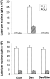
NaV1.5 mRNA accumulation in adult muscle is controlled by activity. Intensity of NaV1.5 labeling per nucleus is nearly fourfold greater at J than XJ nuclei (top panel) in denervated muscle, which is reduced to control levels after electrical stimulation (p < 0.001). Labeling intensity with NaV1.4 probe at J and XJ nuclei is not affected by either denervation or stimulation of adult soleus muscle (bottom panel). Thirty to 40 J clusters and XJ fibers were analyzed for each category.

References

    1. Andreose JS, Fumagalli G, Lømo T. Number of junctional receptors: control by neural and muscular influence in the rat. J Physiol (Lond) 1995;483:397–406. - PMC - PubMed
    1. Bessereau J-L, Stratford-Perricaudet LD, Piette J, Poupon CL, Changeux J-P. In vivo and in vitro analysis of electrical activity-dependent expression of muscle acetylcholine receptor genes using adenovirus. Proc Natl Acad Sci USA. 1994;91:1304–1308. - PMC - PubMed
    1. Boudier JL, Le Treut T, Jover E. Autoradiographic localization of voltage-dependent sodium channels on the mouse neuromuscular junction using 125I-α scorpion toxin. II. Sodium channel distribution on postsynaptic membranes. J Neurosci. 1992;12:454–466. - PMC - PubMed
    1. Bowen DC, Park SJ, Bodine S, Stark JL, Valenzuela DM, Stitt TN, Yancopoulos GD, Lindsay RM, Glass DJ, DiStefano PS. Localization and regulation of Musk at the neuromuscular junction. Dev Biol. 1998;199:309–319. - PubMed
    1. Buonanno A, Fields RD. Gene regulation by patterned electrical activity during neural and skeletal muscle development. Curr Opin Neurobiol. 1999;9:110–120. - PubMed

Publication types

MeSH terms

LinkOut - more resources