. 2001 Nov 1;21(21):8505-13.
doi: 10.1523/JNEUROSCI.21-21-08505.2001.

Phenotypic differentiation during migration of dopaminergic progenitor cells to the olfactory bulb

Affiliations

Phenotypic differentiation during migration of dopaminergic progenitor cells to the olfactory bulb

H Baker et al. J Neurosci. .

Abstract

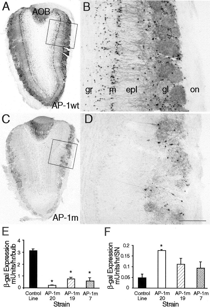
A possible source for transplantable neurons in Parkinson's disease are adult olfactory bulb (OB) dopamine (DA) progenitors that originate in the anterior subventricular zone and reach the OB through the rostral migratory stream. We used adult transgenic mice expressing a lacZ reporter directed by an 8.9 kb tyrosine hydroxylase (TH) promoter to investigate the course of DAergic differentiation. Parallel transgene and intrinsic TH mRNA expression occurred during migration of DA interneurons through the mitral and superficial granule cell layers before these cells reached their final periglomerular position. Differential transgene and calcium-calmodulin-dependent protein kinase IV expression distinguished two nonoverlapping populations of interneurons. Transgenic mice carrying a TH8.9kb/lacZ construct with a mutant AP-1 site demonstrated that this element confers OB DA-specific TH gene regulation. These results indicate that DA phenotypic determination is specific to a subset of mobile OB progenitors.


Figures

Fig. 1.
Fig. 1.
TH protein and mRNA in a mouse OB.A, Immunoreactivity for TH protein is restricted to periglomerular cells of the glomerular (gl) layer and a few cells in the mitral (m) cell layer (arrows,B) and in the accessory olfactory bulb (arrowheads, A). The insetin B shows periglomerular cells at higher magnification;filled arrowheads show a cell and its dendrite ramifying in a glomerulus; the open arrowhead indicates an interglomerular process, presumably an axon. The boxed area delineates the region shown in the inset.C, D, TH mRNA is found in the glomerular, mitral, and superficial granule (gr) cell layers. The RMS does not display either TH protein or mRNA. CTX, Cortex;g, glomerulus. Scale bars: A, C, 650 μm; B, D, 100 μm; inset, 35 μm.
Fig. 2.
Fig. 2.
Tyrosine hydroxylase mRNA demonstrated by nonradioactive in situ hybridization using a digoxigenin-labeled probe and an alkaline phosphatase-conjugated antibody with NBT–BCIP as the chromogen. The low-magnification micrograph (A) shows that label is found only in the olfactory bulb and not in the RMS. The arrowheadindicates the accessory olfactory bulb. B, The higher-magnification micrograph demonstrates that label is heavy in periglomerular cells but lighter in granule cells in the mitral (m) and superficial granule cell (grs) layers (arrows) and absent in the deep granule cell layer (grd). A few presumably migrating cells (arrowhead) exhibiting light label are found in the external plexiform layer (epl).g, Glomerulus. Scale bars: A, 700 μm;B, 100 μm.
Fig. 3.
Fig. 3.
Histochemical (X-Gal) and immunocytochemical (β-gal) demonstration of transgene expression in TH8.9kb/lacZ-expressing mice. Both X-Gal (A) and β-gal (C) label cells in the glomerular (gl), mitral (m), and superficial granule (gr) cell layers but not in the RMS or the cortex (CTX). The granule and mitral cell layers of the accessory olfactory bulb (arrowheads in Aand C) also exhibit transgene expression. Higher-magnification micrographs (B, D) show that the leading processes of labeled cells in the mitral cell layer span the external plexiform layer (epl) and terminate near or within the glomeruli (see also Fig. 4). The high-magnificationinset in D (defined by the white box) shows a labeled cell in the epl (arrowhead) and leading processes (arrow). Scale bars: A, C, 600 μm; B, D, 125 μm;inset, 65 μm.
Fig. 4.
Fig. 4.
Comparison of CaMKII and β-gal immunostaining in the olfactory bulb of TH8.9kb/lacZ-expressing mice. CaMKII staining (A) distinguishes the large mitral cells (arrows) with strong cytoplasmic label and the smaller granule cells (arrowheads) that display only a thin rim of cytoplasmic immunoreactivity. In contrast, β-gal immunostaining (B) is limited to the granule cells of the mitral (m) and granule (gr) cell layers. The processes of these granule cells traverse the external plexiform layer (epl). Scale bars: A, B, 50 μm.
Fig. 5.
Fig. 5.
CaMKIV, β-gal, and TH protein and mRNA expression in the olfactory bulb. A, CaMKIV immunostaining is restricted to deep granule (gr) cells. Low- (B) and high- (C) magnification confocal images of sections double-labeled for β-gal (red) and CaMKIV (green) illustrate the complete separation of the two antigens. Sections immunostained with β-gal (D,red) and TH (E, green) show the large degree of overlap between transgene and TH protein expression in the glomerular (g) region of the olfactory bulb. In the merged image (F), cells containing both antigens are yellow. Gshows that, in contrast to the colocalization of TH and β-gal protein in the glomerular layer, only β-gal immunostaining is observed in the mitral (m) cell layer and few cells exhibit staining in the external plexiform layer (epl). The colocalization of TH mRNA and β-gal protein is shown inH–J. Double-labeled cells appear yellow(arrows in H–J). H′–J′ are higher-magnification images of the mitral cell layer showing that TH mRNA (arrows in I′) but not TH protein is present in granule cells. Scale bars: A, B, 130 μm; C, H–J, 70 μm; D–F, 40 μm; G, 90 μm;H′–J′, 20 μm.
Fig. 6.
Fig. 6.
β-Gal immunostaining contralateral and ipsilateral to unilateral naris closure in TH8.9kb/lacZ-expressing mice. In the olfactory bulb contralateral (c) to naris closure (A), strong fiber and cellular staining are observed in the glomerular (gl) and mitral (m) cell layers. Fibers traverse the external plexiform layer (epl). Ipsilateral (i) to naris closure (B) cellular staining is maintained but fiber staining is dramatically reduced. The inset shows the medial aspects of the olfactory bulbs at low magnification. Letters in the inset indicate the regions illustrated at high magnification inA and B. gr, Granule cell layer; on, olfactory nerve layer. Scale bars: A, B, 100 μm; inset, 650 μm.
Fig. 7.
Fig. 7.
β-Gal immunostaining in wild-type and TH8.9kb AP-1 mutant/lacZ mice. Transgenic mice expressing a transgene with a normal AP-1 construct (A, B) exhibit β-gal immunostaining in the granule (gr), mitral (m), and glomerular (gl) layers. Mice expressing the mutant AP-1 construct show no β-gal staining in the granule and mitral cell layers and reduced staining in the glomerular layer (C, D). The reduction in β-gal expression in the OBs of the TH8.9kb AP-1 mutant/lacZ mice was confirmed by assaying β-gal activity in the OB (E). In the SN (F), β-gal was either the same or higher in mice expressing the mutant construct. Asterisks inE and F indicate significant differences between the mutant lines and the control line. Boxed areas inA and C define areas shown at higher magnification in B and D.epl, External plexiform layer; on, olfactory nerve layer. Scale bars: A, C, 750 μm;B, D, 150 μm.

References

    1. Alvarez-Buylla A, Temple S. Stem cells in the developing and adult nervous system. J Neurobiol. 1998;36:105–110. - PubMed
    1. Anderson S, Mione M, Yun K, Rubenstein JL. Differential origins of neocortical projection and local circuit neurons: role of Dlx genes in neocortical interneuronogenesis. Cereb Cortex. 1999;9:646–654. - PubMed
    1. Anderson SA, Eisenstat DD, Shi L, Rubenstein JL. Interneuron migration from basal forebrain to neocortex: dependence on Dlx genes. Science. 1997a;278:474–476. - PubMed
    1. Anderson SA, Qiu M, Bulfone A, Eisenstat DD, Meneses J, Pedersen R, Rubenstein JL. Mutations of the homeobox genes Dlx-1 and Dlx-2 disrupt the striatal subventricular zone and differentiation of late born striatal neurons. Neuron. 1997b;19:27–37. - PubMed
    1. Baker H. Unilateral, neonatal olfactory deprivation alters tyrosine hydroxylase expression but not aromatic amino acid decarboxylase or GABA immunoreactivity. Neuroscience. 1990;36:761–771. - PubMed

Publication types

MeSH terms