. 2001 Nov 1;21(21):8523-37.
doi: 10.1523/JNEUROSCI.21-21-08523.2001.

Physiological and structural evidence for hippocampal involvement in persistent seizure susceptibility after traumatic brain injury

Affiliations

Physiological and structural evidence for hippocampal involvement in persistent seizure susceptibility after traumatic brain injury

G Golarai et al. J Neurosci. .

Abstract

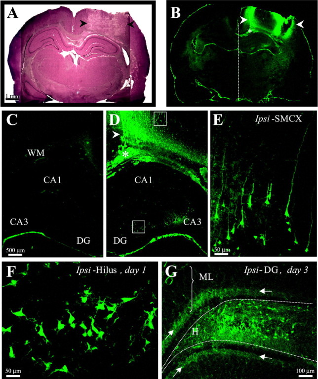
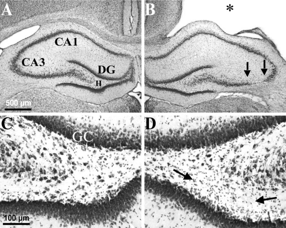
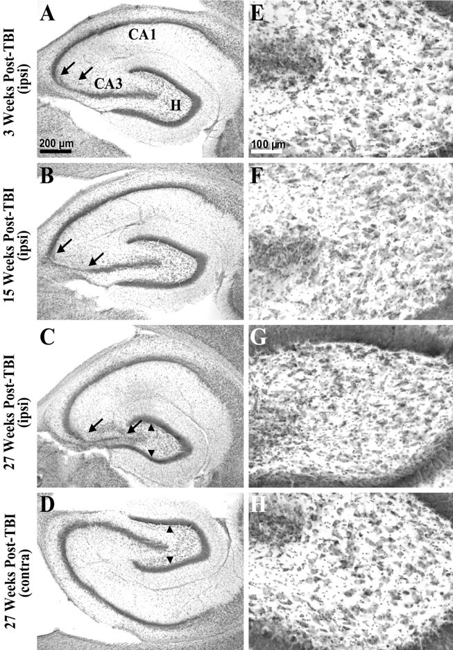
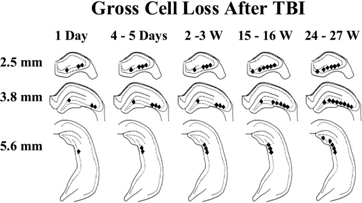
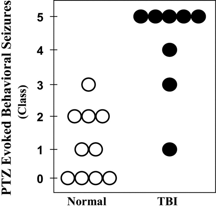
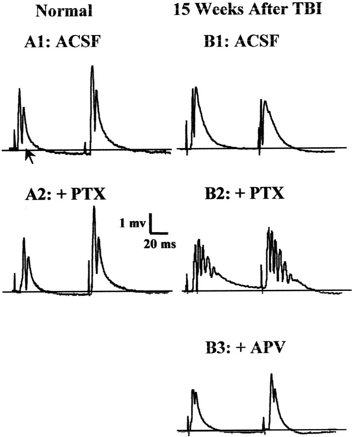
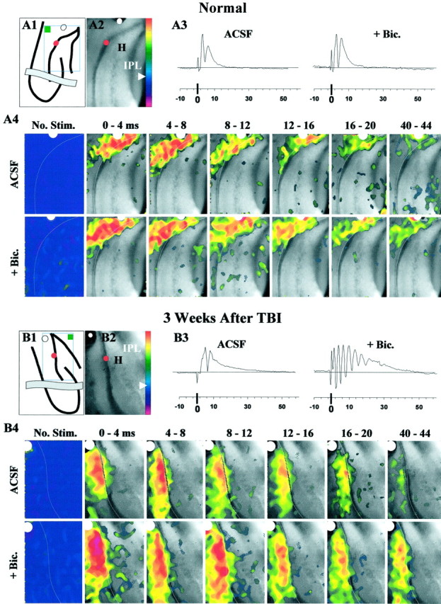
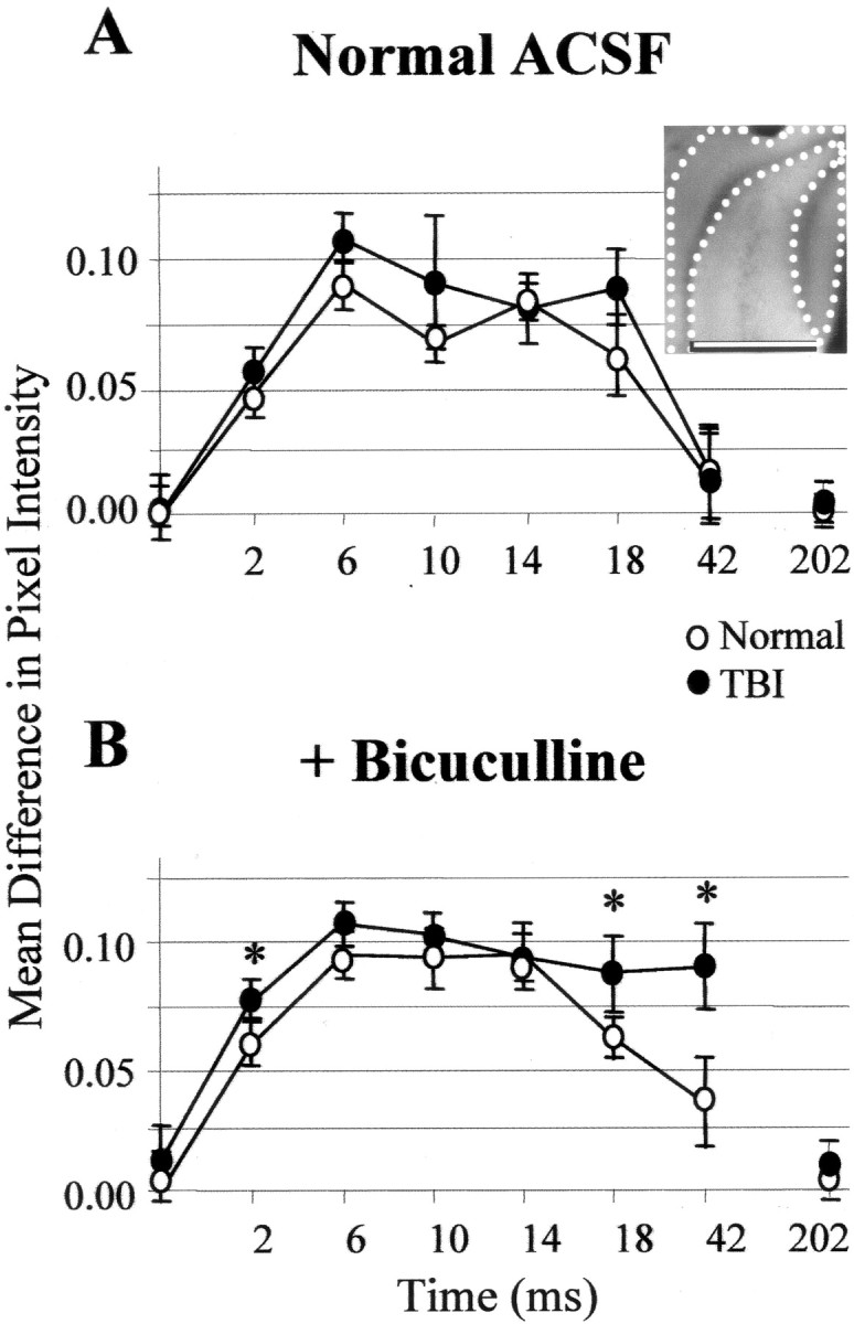
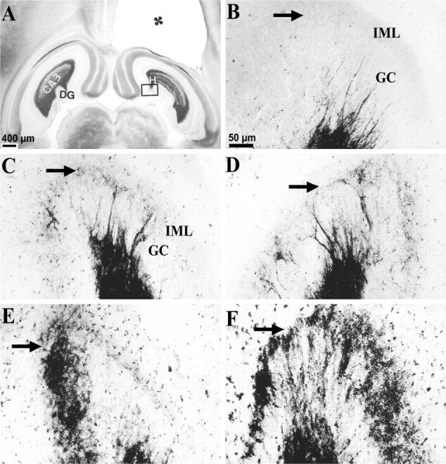
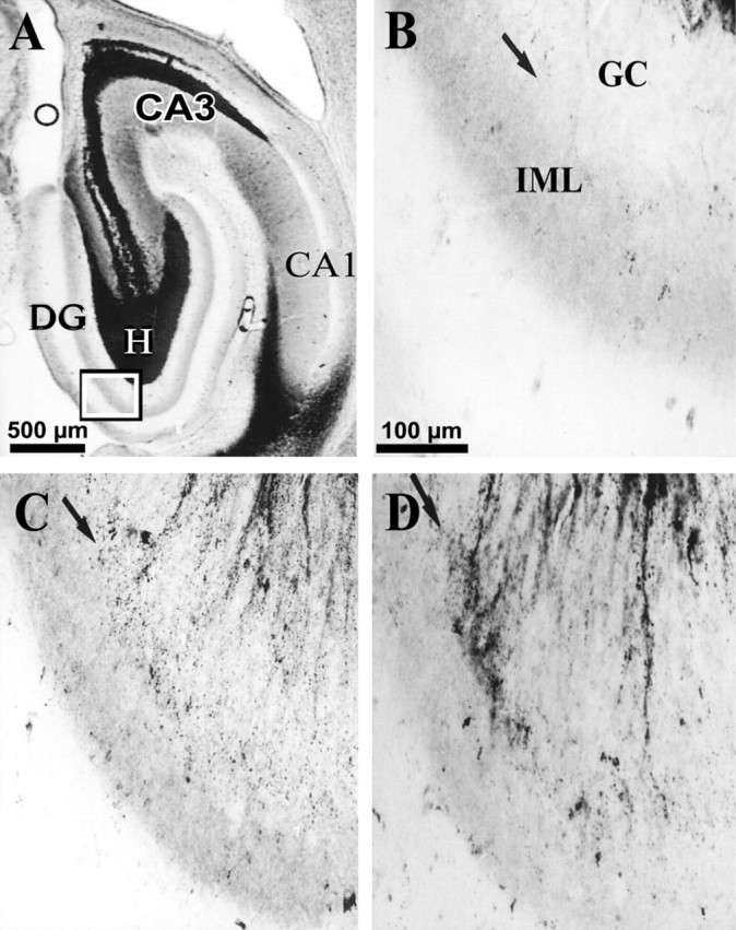
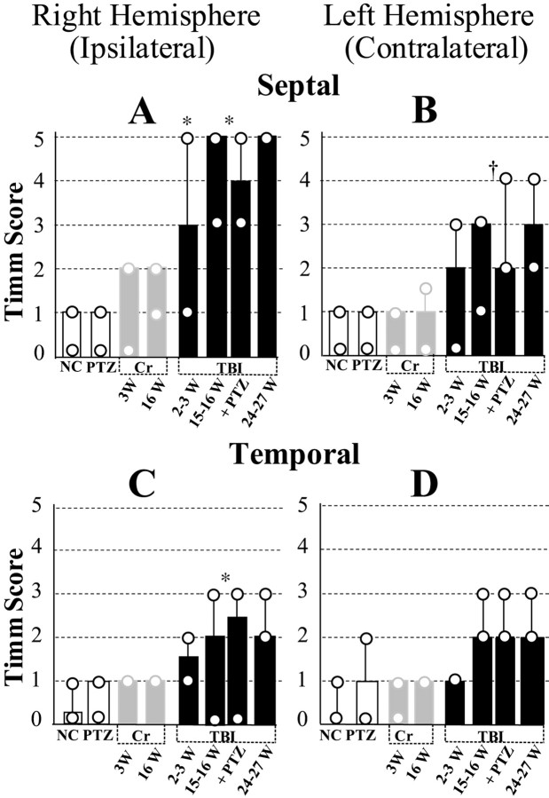
Epilepsy is a common outcome of traumatic brain injury (TBI), but the mechanisms of posttraumatic epileptogenesis are poorly understood. One clue is the occurrence of selective hippocampal cell death after fluid-percussion TBI in rats, consistent with the reported reduction of hippocampal volume bilaterally in humans after TBI and resembling hippocampal sclerosis, a hallmark of temporal-lobe epilepsy. Other features of temporal-lobe epilepsy, such as long-term seizure susceptibility, persistent hyperexcitability in the dentate gyrus (DG), and mossy fiber synaptic reorganization, however, have not been examined after TBI. To determine whether TBI induces these changes, we used a well studied model of TBI by weight drop on somatosensory cortex in adult rats. First, we confirmed an early and selective cell loss in the hilus of the DG and area CA3 of hippocampus, ipsilateral to the impact. Second, we found persistently enhanced susceptibility to pentylenetetrazole-induced convulsions 15 weeks after TBI. Third, by applying GABA(A) antagonists during field-potential and optical recordings in hippocampal slices 3 and 15 weeks after TBI, we unmasked a persistent, abnormal APV-sensitive hyperexcitability that was bilateral and localized to the granule cell and molecular layers of the DG. Finally, using Timm histochemistry, we detected progressive sprouting of mossy fibers into the inner molecular layers of the DG bilaterally 2-27 weeks after TBI. These findings are consistent with the development of posttraumatic epilepsy in an animal model of impact head injury, showing a striking similarity to the enduring behavioral, functional, and structural alterations associated with temporal-lobe epilepsy.


Figures

Fig. 1.
Fig. 1.
Weight-drop TBI induced reproducible damage in the ipsilateral somatosensory cortex and selective cell loss in the ipsilateral hippocampus. A, A cresyl violet-stained coronal section 1 d after TBI shows the impact site in the right somatosensory cortex (arrowheads). This region underwent massive cell loss and by 2 weeks after TBI turned into a fluid-filled cavity (∼30 mm3) spanning cortical layers external to the white matter. B, A Fluoro-Jade-stained coronal section from the same brain asA shows the pattern of cell damage as detected by specific (bright green) Fluoro-Jade labeling of cells 1 d after TBI. Cortical white matter is marked with anasterisk. Arrowheads same asA. One day after TBI, cortical cell damage appeared mainly at the edges of the impact site, with sparse labeling in between. Specific labeling in the CA3 and hilus of the ipsilateral hippocampus is detectable at this low magnification. C, A higher magnification from B shows the cortex and hippocampus contralateral to TBI. Note scarcity of specific labeling contralateral to TBI, compared with D. D, A higher magnification from B shows the cortex and hippocampus ipsilateral to TBI. Arrowhead andasterisk are same as in B. Note the peak of labeling at the medial edge of the impact site (arrowhead) and sparser labeling laterally. Specific labeling is found in ipsilateral CA3 but not CA1. E, A higher magnification of the boxed somatosensory region from D shows labeled cortical neurons with typical pyramidal shapes, some with extended apical and basal processes.F, A higher magnification of the boxedhilar region of the DG from D shows varied shapes of labeled cells, some with dendrite-like processes. G, A Fluoro-Jade-stained coronal section of the ipsilateral DG 3 d after TBI shows labeled cells across the hilus. Arrowspoint to the punctate staining in the dorsal and ventral inner molecular layer (arrows). DG, Dentate gyrus; Ipsi-SMCX, ipsilateral somatosensory cortex; WM, white matter.
Fig. 2.
Fig. 2.
Weight-drop TBI-induced selective, gross cell loss in the ipsilateral hippocampus. A, A cresyl violet-stained coronal section 3 weeks after TBI shows the hippocampus contralateral to TBI with no evidence of macroscopic cell loss.B, From the same brain as A, showing the hippocampus ipsilateral to TBI. Note the TBI-induced cortical cavity (∗). Gross CA3 cell loss is delineated by arrows. There was no obvious cell loss in the CA1 or dentate granule cell layers. C, A higher magnification from Ashows the contralateral hilus with no evidence of gross cell loss after TBI. D, A higher magnification from Bshows the ipsilateral hilus where a subtle loss of large cells can be detected, especially ventrally (between arrows) in this example, as compared with the contralateral side in C.DG, Dentate gyrus; GC, granule cell layer; H, hilus of the dentate gyrus.
Fig. 3.
Fig. 3.
Gross cell loss in the ipsilateral hippocampal CA3 and hilus progressed in temporal regions during the weeks after TBI. A, A cresyl violet-stained horizontal section of the temporal hippocampus from ∼4.5 mm deep with respect to bregma prepared 3 weeks after TBI shows gross cell loss in the ipsilateral CA3 (between arrows). Otherpanels on the left show similarly prepared sections. B, The ipsilateral hippocampus 15 weeks after TBI showed a wider region of gross cell loss in CA3 (between arrows) compared with A.C, The ipsilateral hippocampus 27 weeks after TBI showed a progression of cell loss across the entire CA3. Note atrophy of the ipsilateral hilus, indicated by reduced distance between supragranule and infragranule cell layers (arrowheads) compared withD. D, The contralateral hippocampus 27 weeks after TBI (from the same brain section as C) showed no evidence of gross cell loss.EH show higher magnifications of hilus from AD. E, At 3 weeks after TBI, cell loss was subtle in the ipsilateral hilus in this temporal location. F, At 15 weeks after TBI, cell loss remained subtle in the temporal ipsilateral hilus. G, At 27 weeks after TBI, cell loss and atrophy of the temporal ipsilateral hilus was clearly detectable, although some large hilar neurons remain.H, There was no evidence of gross cell loss in the contralateral hilus 27 weeks after TBI at this temporal location. Scale bars in A and E apply toAD and EH, respectively.
Fig. 4.
Fig. 4.
Selective hippocampal cell loss progressed for weeks after TBI. This summary of the time course and regional distribution of selective cell loss in the hippocampus is based on Fluoro-Jade and cresyl violet staining 1 d (1 Day) to 27 weeks (24–27 W) after TBI, as indicated. Regions of visually discernable cell loss 2.5, 3.8, and 5.6 mm posterior to bregma are marked by filled diamonds. Gross cell loss in the CA3 progressed to temporal regions (i.e., posteriorly) and also from CA3a to CA3c over weeks after TBI. In the hilus, gross cell loss was detected in Fluoro-Jade-stained septal sections by 1 d, in cresyl violet-stained septal sections by 3 weeks, and in temporal hippocampus by 27 weeks after TBI.
Fig. 5.
Fig. 5.
TBI induced a persistent susceptibility to pentylenetetrazole-evoked seizures in vivo (30 mg/kg PTZ, i.p.). Each circle represents a control (open) or a rat 15 weeks after TBI (filled). Convulsions were rated according to a standard scale: no detectable behavioral seizures (class 0); arrest of motion (class I); myoclonic movements (class II); unilateral tonic–clonic seizures (class III); bilateral tonic–clonic seizures (class IV); generalized seizures with loss of postural tone (class V). Generalized seizures were induced in five of eight rats 15 weeks after TBI. In contrast, no generalized seizures were observed in 10 normal rats for 1 hr after PTZ injection.
Fig. 6.
Fig. 6.
TBI induced a persistent hyperexcitability in the DG that was revealed during disinhibition by GABAAantagonists in hippocampal slices 14–15 weeks after TBI.A1, In slices from normal controls in standard ACSF, each of the paired-shock stimuli (60 msec interpulse interval; 100 μsec shock duration) to the perforant path at 0.05 Hz evoked a typical field response in the granule cell layer, consisting of a pEPSP and a single orthodromic population spike (arrow). A2, In the normal DG, addition of GABAA antagonist picrotoxin (PTX, 50 μm) led to a slightly larger amplitude and longer duration population spike, but not to epileptiform activity.B1, In standard ACSF, and 15 weeks after TBI, field-potential responses to perforant-path stimulation lacked epileptiform activity, despite slightly longer than normal pEPSPs. B2, In post-TBI slices in the presence of GABAA antagonist picrotoxin (PTX, 50 μm), shocks to the perforant path induced burst responses consisting of a prolonged depolarization envelope and multiple population spikes. B3, These epileptiform features were eliminated by bath addition of APV (35 μm).
Fig. 7.
Fig. 7.
During simultaneous field and voltage-sensitive optical recordings, perforant-path stimulation evoked burst responses in post-TBI slices in the DG even after isolation from CA3a and CA3b. This activity peaked in the molecular and granule cell layers as shown by the optical data. A1, This line drawing shows the orientation of the hippocampus in a normal slice, an isolating knife cut (gray band), the positions of the stimulating (white dot) and recording (red dot) electrodes, the image frame (open rectangle), and the inactive reference region (filled green box). Color bar was used to display optical signals in pseudocolor as described in Materials and Methods. A2, This representative image of normal DG was acquired during a 4 msec exposure to excitation light. Positions of stimulating (white dot) and recording (red dot) electrodes are marked. A3, Field potentials evoked by single shocks (100 μsec) consisted of a pEPSP and a single population spike in standard ACSF. Addition of bicuculline (+ Bic., 50 μm) did not lead to epileptiform responses. Each trace is an average from 24 consecutive responses evoked every 10–20 sec during collection of the optical data shown in A4 (see Materials and Methods). A4, Panels show color-coded, averaged fluorescence-ratio maps of voltage-sensitive dye signals collected over the specified 4 msec periods after single shocks (at 0 msec). The No Stim. panels show the ratio map with no stimulus, and near zero relative change in optical signals corresponding to pseudocolors indigo topurple. White lines trace the hilar margin of the granule cell layer. Pixels corresponding to pseudocolors below indigo (arrowhead) on thecolor bar in A1 were turned transparent in all panels of A4 except No Stim.ACSF, In standard ACSF in this normal DG slice, perforant-path-evoked depolarization peaked in the molecular and granule cell layers, with minimal spread into the hilus, CA3c, or the opposite blade. + Bic., Bath application of bicuculline (50 μm) did not change the spatial pattern of depolarization. B1, For a rat 3 weeks after TBI,line drawing shows slice orientation as inA1. B2, This representative image of the post-TBI DG is analogous to A2. B3, As inA3, however, addition of bicuculline (50 μm; + Bic) led to burst responses recorded in the granule cell layer. B4, These optical responses correspond to the field potentials in B3.ACSF, In standard ACSF, 3 weeks after TBI, perforant-path stimulation evoked depolarization that peaked in the molecular and granule cell layers of the DG, with minimal spread into the hilus, CA3c, or the opposite blade. + Bic, Bath application of bicuculline (50 μm) increased the amplitude of optical signals at longer latencies (compare corresponding frames at 8–12, 16–20, and40–44 msec with ACSF). During disinhibited burst responses, depolarization peaked in the molecular and granule cell layers, spreading into a limited area in the adjacent infragranular region of the hilus.
Fig. 8.
Fig. 8.
Optically detected depolarization evoked by single shocks to the perforant path was predominantly generated in the granule cell and molecular layers compared with the hilus and CA3c. This difference grew larger at post-shock latencies of 18 and 42 msec (i.e., images recorded 16–20 and 40–44 msec after shock, respectively) during disinhibited burst responses in post-TBI slices relative to normal controls. For a representative slice, the insetshows the regions for which the mean difference in pixel intensity between regions was calculated for inclusion in the group means plotted in A and B. A, For normal (open circles) and TBI (filled circles) slices in ACSF, plotted on the ordinate are the mean pixel intensities in the granule cell and molecular layers (I(GCL + ML); inset: area between dotted lines) minus mean pixel intensities in the hilus and CA3c (I(H + CA3c);inset: area between dotted andsolid lines) for average ratio images at designated times after single shocks. The midpoints of 4 msec recording intervals after single perforant-path shocks are indicated on theabscissa; zero represents no stimulation. The quantity I(GCL + ML)I(H + CA3c) was greater than zero during the period 0 < time < 44, reflecting greater depolarization in the molecular and granule cell layer than in the hilus and CA3c. B, Same as A, but during disinhibition that induced burst responses in post-TBI (but not in normal) slices. These bursts were associated with signals that were larger in the GCL and ML and significantly greater than controls at 18 msec (i.e., 16–20 msec) and 42 msec (i.e., 40–44 msec) after shock (p < 0.05; Bonferroni two-tailedt test). Error bars represent SD.
Fig. 9.
Fig. 9.
MFSR developed in septal DG over weeks after TBI, as detected by Timm staining. Septal MFSR was detected 2 weeks after TBI and became bilaterally more prominent at 16 and 27 weeks after TBI. MFSR is indicated by Timm granules in the SGL, at the border of the granule cell (GC ) and inner molecular layers (IML). A, A Timm-stained horizontal section at ∼3.6 mm deep with respect to bregma is from a rat 3 weeks after TBI. The cavity in the somatosensory cortex (∗) is caused by TBI. Box corresponds to regions shown at higher magnification in BF.B, Timm granules are sparse or absent in the SGL (arrow) of a normal rat. C, At 3 weeks after TBI, an abnormal density of Timm granules was detected in the ipsilateral SGL (arrow) from A.D, At 3 weeks after TBI, clusters of Timm granules were found in the contralateral SGL (arrow) fromA. E, At 16 weeks after TBI, Timm granules formed a confluent band in the ipsilateral SGL (arrow), occasionally extending into the IML.F, At 27 weeks after TBI, Timm granules formed a confluent band in the ipsilateral SGL (arrow), extending into the IML. GC, Granule cell layer; H, hilus; IML, inner molecular layer. Scale bar inB applies toBF.
Fig. 10.
Fig. 10.
Development of MFSR in the ipsilateral temporal hippocampus weeks after TBI, as detected by Timm staining. Temporal posttraumatic MFSR was first detected 16 weeks after TBI and became more prominent by 27 weeks. MFSR is indicated by Timm granules in the SGL at the border of granule cell (GC) and inner molecular layers (IML). A, A Timm-stained horizontal section ∼4.8 mm posterior to bregma is from a rat 16 weeks after TBI. CA3 cell loss is also apparent in this section (open circle). Box corresponds to regions shown at higher magnification in BD.B, In a normal rat, Timm granules were sparse or absent in the SGL (arrow) and IML. C, At 16 weeks after TBI, MFSR was detected as Timm granules distributed along the SGL (arrow). D, At 27 weeks after TBI, MFSR intensified (compared with C), as Timm granules formed larger clusters along the SGL (arrow).DG, Dentate gyrus; GC, granule cell layer; H, hilus; IML, inner molecular layer. Scale bar in B applies toBD.
Fig. 11.
Fig. 11.
Summary of time course and regional distribution of MFSR during the weeks after TBI, based on Timm-stain rating on a five-point scale in standard locations. Posttraumatic MFSR was found in septal and temporal regions of DG in both hemispheres and intensified over time. Across all the time points tested, MFSR was more prominent ipsilateral to TBI in a septal location than contralateral to TBI in a temporal location. A, Ipsilateral septal MFSR was statistically significant 2–3 weeks after TBI versus right hemisphere in normal controls (2–3 W TBI vsNC; *p < 0.05). MFSR intensified from 2 to 15 and 24 weeks after TBI (2–3 W TBI vs pooled data from 15–16 W TBI and 16 W TBI+ PTZ; *p< 0.05). B, Contralateral septal MFSR reached statistical significance 15–16 weeks after TBI (pooled data vs left hemisphere NC; p < 0.01) and persisted beyond 24 weeks after TBI. C, Ipsilateral temporal MFSR reached statistical significance 15–16 weeks after TBI (pooled data vs right hemisphere NC; *p < 0.05) and persisted beyond 24 weeks after TBI. D, Contralateral temporal MFSR was detected at 15–16 weeks after TBI and persisted beyond 24 weeks after TBI.NC, Normal control, n = 9;PTZ, PTZ controls (1 d after 1 injection of PTZ, 30 mg/kg, i.p.), n = 9; 3W Cr, 3 weeks after craniotomy alone (no weight drop), n = 5;16W Cr, 16 weeks after craniotomy alone,n = 3; 2–3 W TBI, 2–3 weeks after TBI,n = 9; 15–16 W TBI: 15–16 weeks after TBI,n = 6; 16 W TBI + PTZ, 16 weeks after TBI and 1 d after one injection of PTZ (30 mg/kg, i.p),n = 7; 24–27 W TBI, 24–27 weeks after TBI, n = 3.

References

    1. Aamodt SM, Constantine-Paton AU. The role of neural activity in synaptic development and its implications for adult brain function. Adv Neurol. 1999;79:133–144. - PubMed
    1. Annegers JF, Rocca WA, Hauser WA. Causes of epilepsy: contributions of the Rochester epidemiology project. Mayo Clin Proc. 1996;71:570–575. - PubMed
    1. Annegers JF, Hauser WA, Coan SP, Rocca WA. A population-based study of seizures after traumatic brain injuries. N Engl J Med. 1998;338:20–24. - PubMed
    1. Asikainen I, Kaste M, Sarna S. Early and late posttraumatic seizures in traumatic brain injury rehabilitation patients: brain injury factors causing late seizures and influence of seizures on long-term outcome. Epilepsia. 1999;40:584–589. - PubMed
    1. Babb TL. Axonal growth and neosynaptogenesis in human and experimental hippocampal epilepsy. Adv Neurol. 1997;72:45–51. - PubMed

Publication types

MeSH terms