. 2001 Nov 1;21(21):8538-47.
doi: 10.1523/JNEUROSCI.21-21-08538.2001.

Semaphorin 3A elicits stage-dependent collapse, turning, and branching in Xenopus retinal growth cones

Affiliations

Semaphorin 3A elicits stage-dependent collapse, turning, and branching in Xenopus retinal growth cones

D S Campbell et al. J Neurosci. .

Abstract

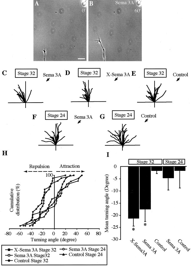
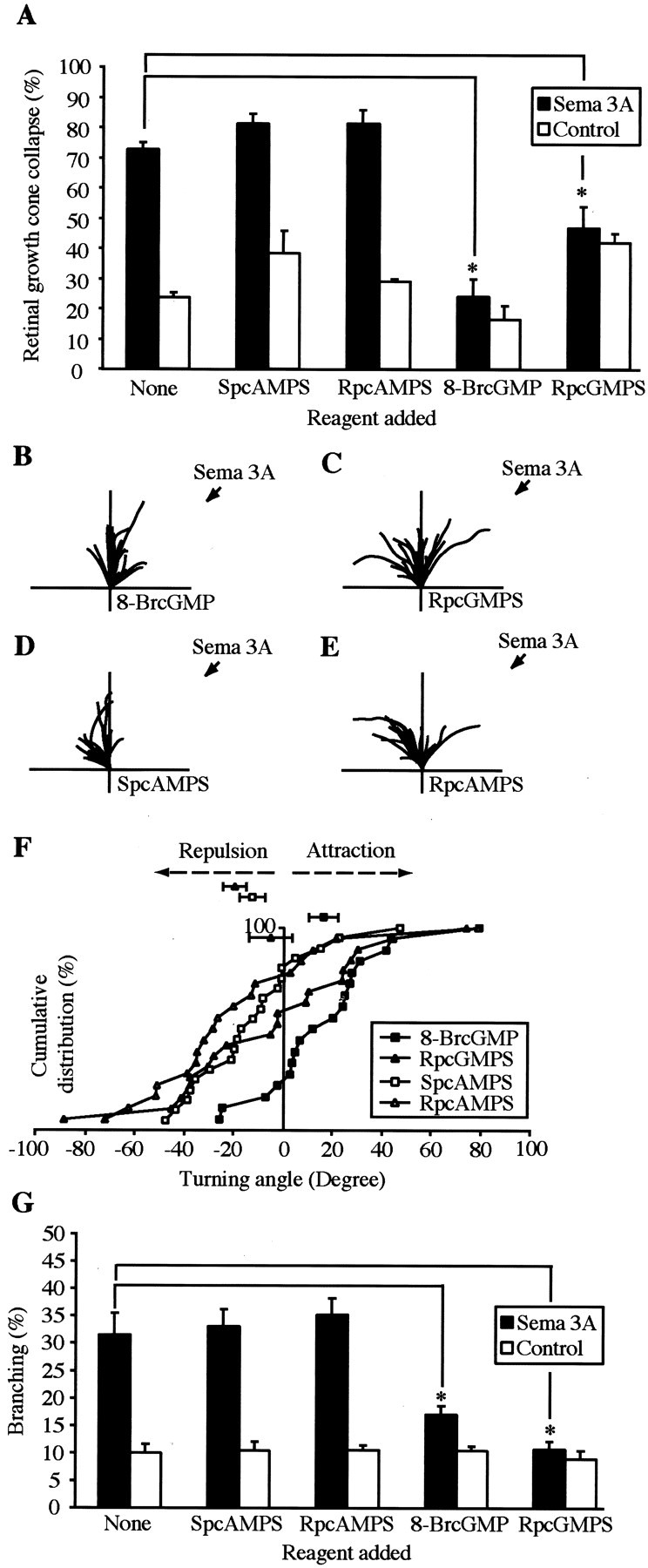
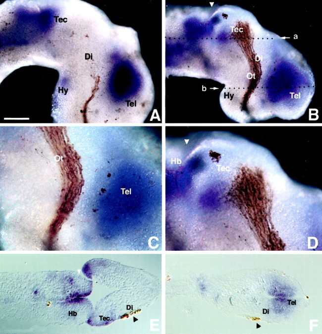
The semaphorin receptor, neuropilin-1 (NP-1), was first identified in Xenopus as the A5 antigen and is expressed abundantly in developing retinal ganglion cells (RGCs). Here we show that growth cones acquire responsiveness to semaphorin 3A (Sema 3A) with age and that the onset of responsiveness correlates with the appearance of NP-1 immunoreactivity. Growth cones from "old" (stage 35/36) retinal explants collapse rapidly (5-10 min) in response to Sema 3A and turn away from a gradient of Sema 3A, whereas "young" growth cones (stage 24) are insensitive to Sema 3A. Moreover, transfection of full-length NP-1 into young neurons confers premature Sema 3A sensitivity. When young neurons are aged in culture they develop Sema 3A sensitivity in parallel with those in vivo, suggesting that an intrinsic mechanism of NP-1 regulation mediates this age-dependent change. Sema 3A-induced collapse is transient, and after recovery approximately 30% of growth cones extend new branches within 1 hr, implicating Sema 3A as a branching factor. Pharmacological inhibitors were used to investigate whether these three Sema 3A-induced behaviors (collapse, turning, and branching) use distinct second messenger signaling pathways. All three behaviors were found to be mediated via cGMP. In situ hybridization shows that Sema 3A is expressed in the tectum and at the anterior boundary of the optic tract where axons bend caudally, suggesting that Sema 3A/NP-1 interactions play a role in guiding axons in the optic tract and in stimulating terminal branching in the tectum.


Figures

Fig. 1.
Fig. 1.
Sema 3A induces rapid and transient collapse of retinal growth cones. A, The morphology of a retinal growth cone before the bath application of Sema 3A; B, a collapsed retinal growth cone 10 min after Sema 3A application;C, recovery of the growth cone after 60 min.D, Sema 3A induces the transient collapse of 24 hr stage 35/36 growth cones. E, Sema 3A and X-Sema 3A-induced collapse are dose dependent. *p < 0.05; Mann–Whitney U test. Scale bar (shown inA): AC, 10 μm.
Fig. 2.
Fig. 2.
Retinal growth cones gain Sema 3A responsiveness with age. A, Sema 3A-induced collapse for 24 hr stages 24, 28, 35/36, and 37/38 retinal growth cones. Stages 35/36 and 37/38 growth cones show significant increases in collapse compared with stages 24 and 28, *p < 0.01; ANOVA.B, Sema 3A-induced collapse for 6, 24, or 48 hr stage 24 growth cones. Sema 3A-induced stage 24 growth cone collapse is significantly increased when cultured for 48 hr. *p< 0.05; Mann–Whitney U test. NP-1 and plexin expression increase in stage 24 plated retinal growth cones with duration in culture. Shown are representative photographs of NP-1 (CE) and plexin (GI) expression by growth cones using the A5 and B2 antibodies and a Cy3-conjugated secondary. Levels of NP-1 (F) and plexin (J) expression were quantified comparing percentage fluorescence intensity relative to control (without primary antibody). *p< 0.05; Student's t test (C,G). Sema 3A-induced collapse correlates with NP-1 expression. A 6 hr stage 24 growth cone (K) does not collapse in response to the bath application of Sema 3A (L) and does not express NP-1 (M). A 24 hr stage 35/36 growth cone (N) collapses in response to bath application of Sema 3A (O) and expresses high levels of NP-1 (P).
Fig. 3.
Fig. 3.
Precocious expression of NP-1 is sufficient to confer Sema 3A responsiveness on young retinal growth cones.A, The NP-1 antibody (AN-1) inhibits Sema 3A-induced collapse. B, A 6 hr stage 24 retinal growth cone expressing NP-1-myc. C, A collapsed growth cone expressing NP-1-myc in the presence of Sema 3A. Scale bar, 10 μm.D, Sema 3A induces the collapse of 6 hr stage 24 retinal growth cones expressing NP-1-myc. *p < 0.05; Mann–Whitney U test.
Fig. 4.
Fig. 4.
Sema 3A elicits repulsive turning in old retinal growth cones. A, A 16–26 hr stage 32 retinal growth cone before being exposed to a gradient of Sema 3A. Scale bar, 10 μm.B, After 60 min the growth cone is repelled by a gradient of Sema 3A. Traces depict the trajectories of 16–26 hr stage 32 neurites in the presence of a Sema 3A gradient applied at the black arrow (C) or X-Sema 3A gradient (D) compared with a gradient of control supernatant (Control, E).Traces depict the trajectories of 6–12 hr stage 24 retinal neurites in the presence of a gradient of Sema 3A that does not induce repulsive turning (F) and control (G). H, Cumulative frequency graph showing the distribution of turning angles for stages 32 and 24 retinal growth cones, Sema 3A, and control. Sema 3A and X-Sema 3A are repulsive to growth cones from stage 32 plated retinal explants; i.e., most of the turning angles are negative relative to control turning angles and the turning angles for a gradient of Sema 3A on stage 24 growth cones and control. I, Mean turning, Sema 3A, and X-Sema 3A induce significant repulsion of stage 32 retinal growth cones. *p < 0.05; Kolmogorov–Smirnov test.
Fig. 5.
Fig. 5.
Sema 3A elicits branching after recovery from collapse. A, A 24 hr stage 35/36 retinal growth cone before the application of Sema 3A. B, The same growth exhibiting collapsed morphology after 10 min. C, After 30 min the growth cone has begun to branch. D, After 60 min the branch remains. Scale bar, 10 μm. E, Sema 3A induces the branching of retinal growth cones. Cumulative branching in the most distal 100 μm of neurite growth illustrates that the majority of the branching occurs within approximately the first 35 μm, the mean neurite growth in 1 hr. *p < 0.05; Mann–Whitney U test.
Fig. 6.
Fig. 6.
Pharmacological perturbation of cGMP signaling modulates the responses to Sema 3A. Pharmacological perturbation of cGMP signaling but not cAMP signaling via the activation or inhibition of protein kinase G significantly reduces 24 hr stage 35/36 Sema 3A-induced growth cone collapse (A). *p < 0.05; Mann–Whitney U test.Traces depict the trajectories of 16–26 hr stage 32 retinal neurites in the presence of pharmacological modulators of cGMP and cAMP signaling (BE) in the medium and a directional source of Sema 3A. B, The activation of PKG with 100 μm 8-BrcGMP converts Sema 3A-induced repulsion to attraction. p < 0.01; Kolmogorov–Smirnov test.C, Inhibition of PKG with 10 μm RpcGMPS abolishes Sema 3A-induced repulsion, leading to a heterogeneous turning response. D, E, Activation or inhibition of PKA with 20 μm SpcAMPS or RpAMPS does not significantly affect Sema 3A-induced repulsion. F, Cumulative frequency graph showing the turning angles to Sema 3A. In the presence of 100 μm 8-BrcGMP, the curve is shifted to theright. With 10 μm RpcGMPS, approximately equal numbers of angles lie on either side, and with 20 μm SpcAMPS or RpcAMPS, most turning angles lie to theleft, indicating repulsion. G, Activation or inhibition of PKG inhibits Sema 3A-induced branching. *p < 0.05; Mann–Whitney Utest.
Fig. 7.
Fig. 7.
Sema 3A expression in the developing optic pathway. Shown are whole-mount lateral views of stage 33/34 (A) and stage 41 (BD) Xenopus brains in which the RGCs have been anterogradely filled with HRP and visualized with DAB. Sema 3A is highly expressed in the telencephalon, hindbrain, and posterior tectum but not in the optic tract (A,B). Shown is magnified view of Sema 3A expression in the diencephalon (C) and posterior tectum (D) illustrating its proximity to the RGC axons. Shown are horizontal paraffin sections at the level of the tectum (E) and telencephalon (F). The level of the sections in E and F is denoted by the white arrow in B, labeleda and b, respectively. Di, Diencephalon; Hb, hindbrain; Hy, hypothalamus; Ot, optic tract; Tec, tectum; Tel, telencephalon. White arrowheads indicate the midbrain/hindbrain boundary.Black arrowheads highlight the HRP-filled RGC axons. Scale bar (shown in A): A,B, E, F, 100 μm;C, D, 50 μm. Anterior is to theright.

References

    1. Acebes A, Ferrus A. Cellular and molecular features of axon collaterals and dendrites. Trends Neurosci. 2000;23:557–565. - PubMed
    1. Bagnard D, Lohrum M, Uziel D, Puschel AW, Bolz J. Semaphorins act as attractive and repulsive guidance signals during the development of cortical projections. Development. 1998;125:5043–5053. - PubMed
    1. Brose K, Tessier-Lavigne M. Slit proteins: key regulators of axon guidance, axonal branching, and cell migration. Curr Opin Neurobiol. 2000;10:95–102. - PubMed
    1. Castellani V, Yue Y, Gao PP, Zhou R, Bolz J. Dual action of a ligand for Eph receptor tyrosine kinases on specific populations of axons during the development of cortical circuits. J Neurosci. 1998;18:4663–4672. - PMC - PubMed
    1. Chien CB, Harris WA. Axonal guidance from retina to tectum in embryonic Xenopus. Curr Top Dev Biol. 1994;29:135–169. - PubMed

Publication types

MeSH terms