. 2001 Nov 1;21(21):8572-85.
doi: 10.1523/JNEUROSCI.21-21-08572.2001.

Transforming growth factor beta (TGFbeta) mediates Schwann cell death in vitro and in vivo: examination of c-Jun activation, interactions with survival signals, and the relationship of TGFbeta-mediated death to Schwann cell differentiation

Affiliations

Transforming growth factor beta (TGFbeta) mediates Schwann cell death in vitro and in vivo: examination of c-Jun activation, interactions with survival signals, and the relationship of TGFbeta-mediated death to Schwann cell differentiation

D B Parkinson et al. J Neurosci. .

Abstract

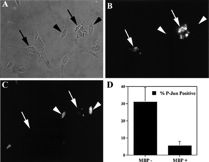
In some situations, cell death in the nervous system is controlled by an interplay between survival factors and negative survival signals that actively induce apoptosis. The present work indicates that the survival of Schwann cells is regulated by such a dual mechanism involving the negative survival signal transforming growth factor beta (TGFbeta), a family of growth factors that is present in the Schwann cells themselves. We analyze the interactions between this putative autocrine death signal and previously defined paracrine and autocrine survival signals and show that expression of a dominant negative c-Jun inhibits TGFbeta-induced apoptosis. This and other findings pinpoint activation of c-Jun as a key downstream event in TGFbeta-induced Schwann cell death. The ability of TGFbeta to kill Schwann cells, like normal Schwann cell death in vivo, is under a strong developmental regulation, and we show that the decreasing ability of TGFbeta to kill older cells is attributable to a decreasing ability of TGFbeta to phosphorylate c-Jun in more differentiated cells.


Figures

Fig. 1.
Fig. 1.
TGFβ induces apoptosis in Schwann cellsin vitro. AC, Death of Schwann cells at high density on polyornithine induced by TGFβ (2 ng/ml) as measured by criteria of cell morphology (A), nuclear condensation viewed by Hoechst stain (B), and TUNEL labeling (C). Arrows indicate live cells with extended processes with nuclei that are round and TUNEL negative.Arrowheads show dying or dead cells that are rounded up, have condensed nuclei, and are TUNEL positive. D, Counts of live and dead cells in control (−) and TGFβ1 (2 ng/ml)-treated (+) cultures of Schwann cells at low density (LD/PO) and high density (HD/PO) on polyornithine, and high density on laminin substrate (HD/LAM). E,F, Activation of caspase 3 in dying Schwann cells treated with TGFβ1 (2 ng/ml). Arrows indicate live cells as viewed by phase contrast (E) that are not labeled for active caspase 3 (F). Examples of dying/dead cells, which are labeled for active caspase-3, are indicated with arrowheads.
Fig. 2.
Fig. 2.
In vitro survival assays. TGFβ-induced cell death is not blocked by high cell density or by NRGβ alone. A, TGFβ1 causes death of Schwann cells in a dose-dependent manner. Schwann cells plated at low density (300 cells per coverslip) on polyornithine-coated coverslips were treated with increasing concentrations of TGFβ1, and survival was assessed after 24 hr. B, Cell death caused by TGFβ1 is density independent. Survival of Schwann cells plated on polyornithine at different densities as indicated and treated with TGFβ1 (2 ng/ml) for 24 hr is shown. C, Dose-dependent TGFβ1-induced death occurs even in high-density cultures (3000 cells per coverslip) on a laminin substrate. D, In low-density cultures (300 cells per coverslip), NRGβ is relatively ineffective at inhibiting TGFβ1-induced death. Schwann cells were treated with TGFβ1 (2 ng/ml) in the presence of increasing concentrations of NRGβ; survival was assessed after 24 hr. Compare this curve with that shown in Figure3A.
Fig. 3.
Fig. 3.
In vitro survival assays. TGFβ-induced cell death is blocked by a combination of NRGβ and autocrine signals. A, In high-density Schwann cell cultures (3000 cells per coverslip), NRGβ inhibits TGFβ1 (2 ng/ml)-induced apoptosis in a dose-dependent manner. B, In sister cultures, application of NRGβ at 10 ng/ml in the presence of increasing amounts of TGFβ1 (TGFB+NRG) inhibits apoptosis. Shown also are data for TGFβ1 alone (TGFB).C, In the presence of TGFβ1 (2 ng/ml) in low-density cultures (300 cells per coverslip), NRGβ (NRG, 10 ng/ml) alone, or a combination of IGF-2 (1.6 ng/ml), NT3 (0.8 ng/ml), and PDGF-BB (0.8 ng/ml) (Auto) alone, inhibits TGFβ1-induced apoptosis only partially, whereas application of both (NRG+Auto.) blocks TGFβ1-induced death.
Fig. 4.
Fig. 4.
A, Nerve transection in newborn rat causes an upregulation of TGFβ1 mRNA and downregulation of TGFβ3 mRNA. Shown is semiquantitative PCR measurement of levels of mRNA for TGFβ1 and TGFβ3 in newborn rat sciatic nerve (NB), 1 d (1d NB-TS), and 3 d (3d NB-TS) after transection, and from postnatal day 3 (P3) rat. B, Measurement of TGFβ1 protein levels in intact and transected newborn nerve. Shown is Western blot of protein from newborn (NB), P2, and newborn rat sciatic nerve 2 d after transection (2d NB-TS).
Fig. 5.
Fig. 5.
TUNEL analysis of newborn rat sciatic nerve after transection. Shown are representative fields from sections stained with Hoechst dye to reveal nuclei (A, C,E) and TUNEL stained to reveal apoptotic nuclei (B, D, F).A and B show control untransected nerve,C and D show sciatic nerve 24 hr after transection, and E and F show transected nerve 24 hr after transection with injection of pan-TGFβ blocking antibody. Arrows point to examples of individual TUNEL-labeled nuclei.
Fig. 6.
Fig. 6.
In vitro survival assays. The ability of TGFβ1 to cause apoptosis of Schwann cells is developmentally regulated. A, Survival of Schwann cells from newborn (NB), postnatal day 4 (P4), and P8 rats plated at high density (3000 cells per coverslip) with increasing concentrations of TGFβ1. Note that the ability of TGFβ to kill Schwann cells decreases as the cells get older. B, Survival assay for Schwann cells showing percentage of MBP-positive cells (MBP+) and MBP-negative cells (MBP−) from P4 animals in cultures treated with TGFβ1 (5 ng/ml) in a 24 hr assay. Note that the percentage survival of MBP-positive cells (cells that were myelinating in vivo) is relatively unaffected by TGFβ1.
Fig. 7.
Fig. 7.
Expression of type I TGFβ receptors ALK1, ALK2, and ALK5 in sciatic nerve during development. Semiquantitative RT-PCR measurement of ALK1, ALK2, and ALK5 from E14 through to adult (Ad.) in rat sciatic nerve. Note marked downregulation of ALK2 mRNA in postnatal nerves.
Fig. 8.
Fig. 8.
Addition of TGFβ1 to newborn rat Schwann cellsin vitro causes serine-63 phosphorylation of c-Jun, activation of AP1-dependent transcriptional activity, and induction of cyclin D1 mRNA. A, C, andE show immunopanned newborn rat Schwann cells stained with Hoechst dye to reveal the nuclei, and B,D, and F show Schwann cells immunolabeled with antibody specific for the serine-63-phosphorylated form of c-Jun.A and B show control untreated cells with low levels of phosphorylated c-Jun. C andD show Schwann cells treated for 1 hr with TGFβ1 (2 ng/ml). Note substantial serine-63 phosphorylation of c-Jun.E and F are
Fig. 9.
Fig. 9.
Overexpression of dominant negative c-Jun inhibits TGFβ1-induced apoptosis in Schwann cells. A,B, Immunolabeling of Schwann cells with anti-FLAG antibody 48 hr after addition of adenoviral supernatants before TGFβ1 addition shows expression of FLAG-tagged dominant negative c-Jun (FLAGΔ169-Jun) in >90% of Schwann cell nuclei. Ashows Hoechst stain of nuclei; B shows immunofluorescence with FLAG antibody. C, In vitro survival assays of Schwann cells infected with control virus (Lac-Z) or with dominant negative c-Jun (FLAG169-Jun). The cells were exposed to medium alone or to medium containing 10 or 20 ng/ml TGFβ1 for 24 hr. Schwann cells treated with UV light, a known activator of c-Jun phosphorylation, as positive control. G andH show immunolabeling in control (G) and TGFβ1-treated (H) (2 ng/ml for 1 hr) Schwann cells to show that c-Jun protein levels remain unaltered at this time point after TGFβ1 addition. c-Jun mRNA as assayed by semiquantitative RT-PCR (I) also remains unaltered at this time. J shows RT-PCR data demonstrating TGFβ1 (2 ng/ml) induction of cyclin D1 mRNA; amplification using primers specific for 18S rRNA indicates equal input of cDNA into PCR assays.K shows that TGFβ1 causes strong induction of AP1-dependent transcription in Schwann cells. Schwann cells were cotransfected with an AP1-dependent CAT reporter construct together with pCH110 lac-Z plasmid. Thirty hours after transfection in the absence (Control) or presence of 5 ng/ml of TGFβ1 (+TGFB1), lysates were assayed for CAT activity.
Fig. 10.
Fig. 10.
Overexpression of v-Jun causes apoptosis of Schwann cells in serum-free medium. Schwann cells infected with either LexA control (Control) or LexA-vJun (v-Jun) were plated at high density (5000 cells per coverslip). The medium of the cells was then changed to supplemented defined medium, and the number of surviving cells was counted after 48 hr.
Fig. 11.
Fig. 11.
Phosphorylation of c-Jun in Schwann cells by TGFβ1 occurs mostly in MBP-negative cells. Immunopanned Schwann cells from P4 rats were treated for 1 hr with TGFβ1 (2 ng/ml) and double immunolabeled with antibodies to MBP (B) and ser-63 phospho c-Jun (C). After addition of TGFβ1, cells indicated by arrowheads in the phase-contrast micrograph (A) are shown to be negative for MBP (B) but positive for ser-63- phosphorylated c-Jun (C). The two MBP-positive cells in this field (arrows) are negative for phosphorylated c-Jun. Note the typical vesicular appearance of MBP in Schwann cells in culture after loss of axonal contact. Dquantifies this effect, illustrating the percentage of MBP-negative (MBP−) and MBP-positive (MBP+) Schwann cells that are also positive for the phosphorylated serine-63 form of c-Jun in this assay.
Fig. 12.
Fig. 12.
ICE protease inhibitor Z-VAD.fmk inhibits TGFβ1-induced apoptosis in Schwann cells. The graph shows 24 hr survival assays for immunopanned newborn rat Schwann cells plated at low density (300 cells per coverslip) on polyornithine-coated coverslips. Cells were maintained in defined medium alone (DM), defined medium supplemented with NRGβ (10 ng/ml) (NRG), Z-VAD.fmk (100 μm) (Z-VAD), TGFβ1 (2 ng/ml) (TGFB), or TGFβ1 plus Z-VAD.fmk (TGFB+Z-VAD).

Similar articles

Cited by

References

    1. Albanese C, Johnson J, Watanabe G, Eklund N, Vu D, Arnold A, Pestell RG. Transforming p21ras mutants and c-Ets-2 activate the cyclin D1 promoter through distinguishable regions. J Biol Chem. 1995;270:23589–23597. - PubMed
    1. Bader AG, Hartl M, Bister K. Conditional cell transformation by doxycycline-controlled expression of the ASN17 v-Jun allele. Virology. 2000;270:98–110. - PubMed
    1. Bakiri L, Lallemand D, Bossy-Wetzel E, Yaniv M. Cell cycle-dependent variations in c-Jun and JunB phosphorylation: a role in the control of cyclin D1 expression. EMBO J. 2000;19:2056–2068. - PMC - PubMed
    1. Berkner KL. Development of adenovirus vectors for the expression of heterologous genes. Biotechniques. 1998;6:616–629. - PubMed
    1. Black EJ, Catling AD, Woodgett JR, Kilbey A, Gillespie DA. Transcriptional activation by the v-Jun oncoprotein is independent of positive regulatory phosphorylation. Oncogene. 1994;9:2363–2368. - PubMed

Publication types

MeSH terms

LinkOut - more resources