. 2001 Nov 1;21(21):8602-15.
doi: 10.1523/JNEUROSCI.21-21-08602.2001.

Neurotrophin-4 deficient mice have a loss of vagal intraganglionic mechanoreceptors from the small intestine and a disruption of short-term satiety

Affiliations

Neurotrophin-4 deficient mice have a loss of vagal intraganglionic mechanoreceptors from the small intestine and a disruption of short-term satiety

E A Fox et al. J Neurosci. .

Abstract

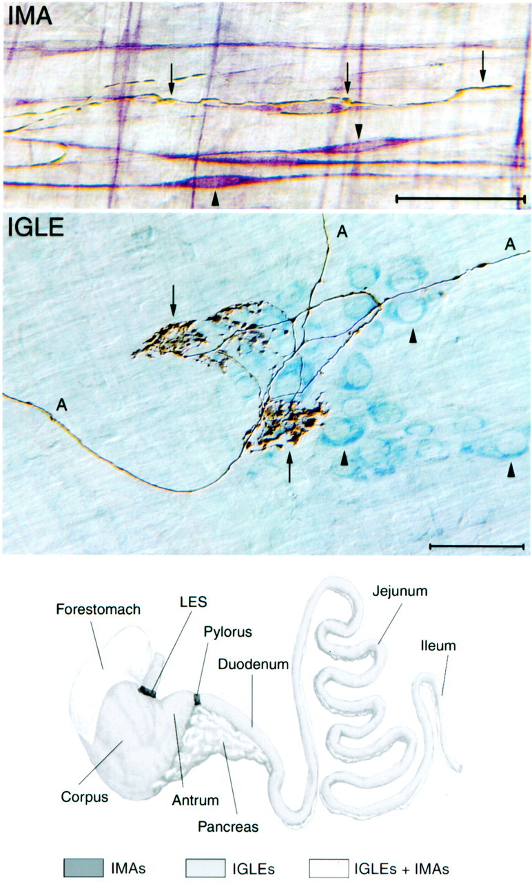
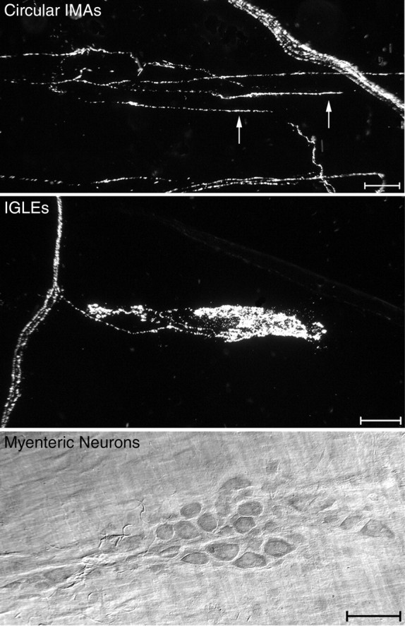
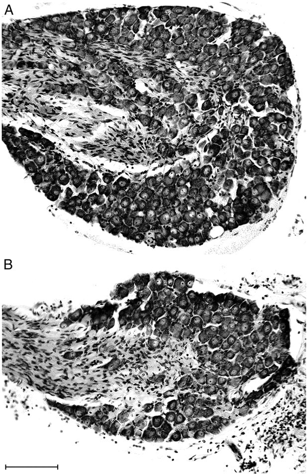
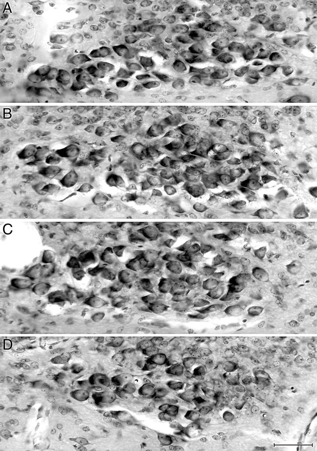
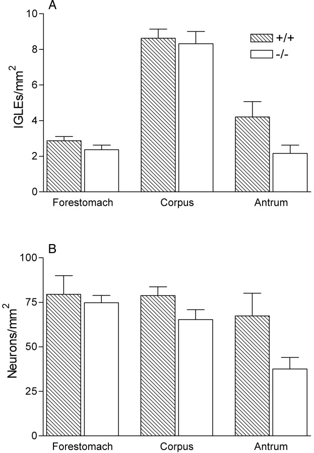
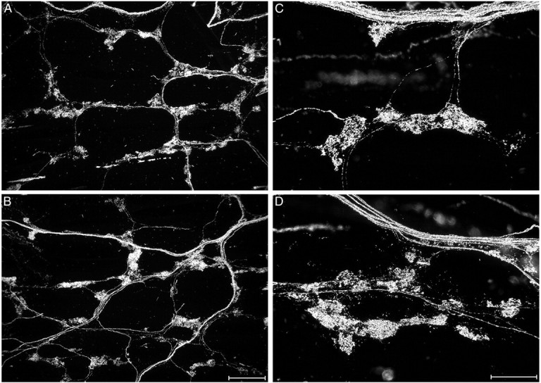
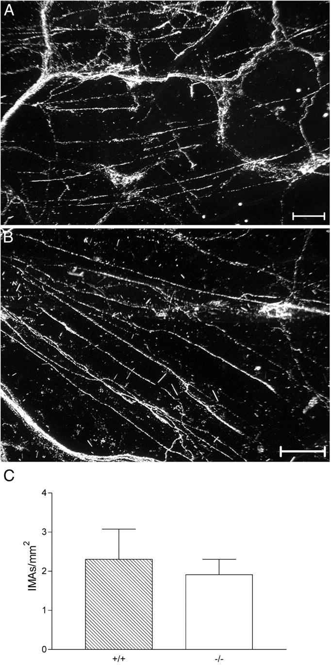
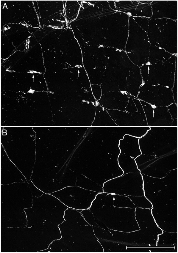
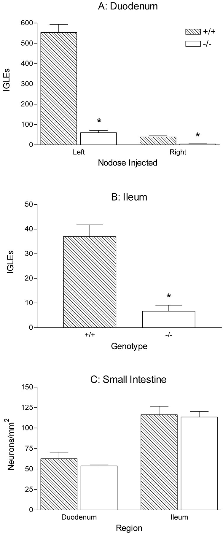
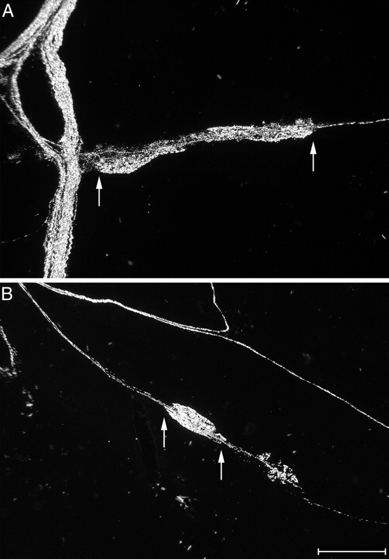
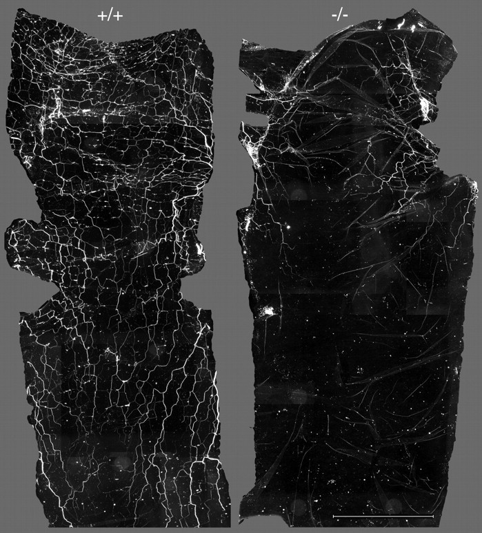
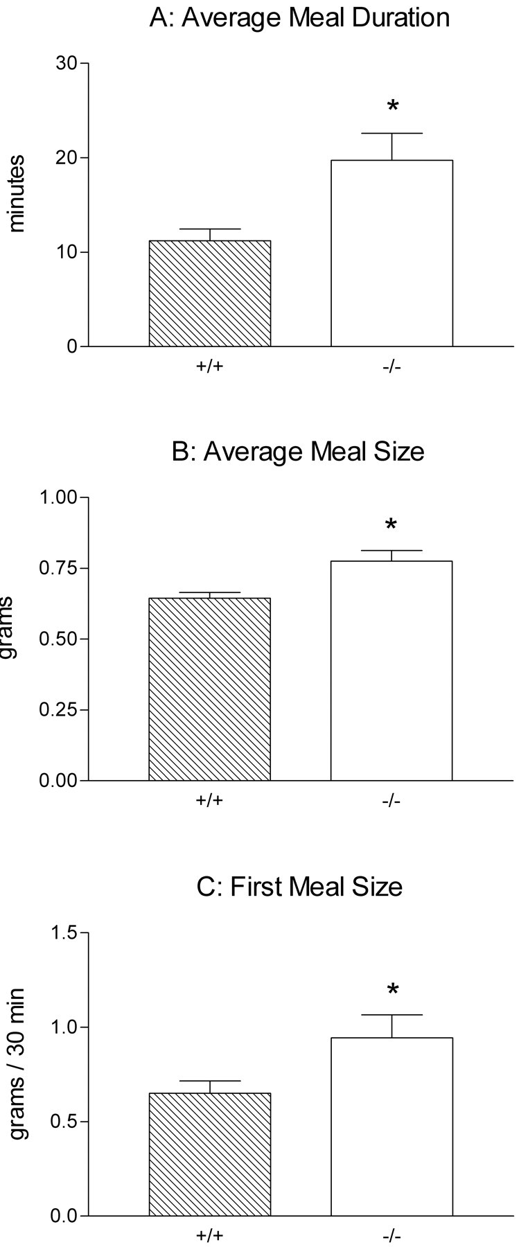
Intraganglionic laminar endings (IGLEs) and intramuscular arrays (IMAs) are the two putative mechanoreceptors that the vagus nerve supplies to gastrointestinal smooth muscle. To examine whether neurotrophin-4 (NT-4)-deficient mice, which have only 45% of the normal number of nodose ganglion neurons, exhibit selective losses of these endings and potentially provide a model for assessing their functional roles, we inventoried IGLEs and IMAs in the gut wall. Vagal afferents were labeled by nodose ganglion injections of wheat germ agglutinin-horseradish peroxidase, and a standardized sampling protocol was used to map the terminals in the stomach, duodenum, and ileum. NT-4 mutants had a substantial organ-specific reduction of IGLEs; whereas the morphologies and densities of both IGLEs and IMAs in the stomach were similar to wild-type patterns, IGLEs were largely absent in the small intestine (90 and 81% losses in duodenum and ileum, respectively). Meal pattern analyses revealed that NT-4 mutants had increased meal durations with solid food and increased meal sizes with liquid food. However, daily total food intake and body weight remained normal because of compensatory changes in other meal parameters. These findings indicate that NT-4 knock-out mice have a selective vagal afferent loss and suggest that intestinal IGLEs (1) may participate in short-term satiety, probably by conveying feedback about intestinal distension or transit to the brain, (2) are not essential for long-term control of feeding and body weight, and (3) play different roles in regulation of solid and liquid diet intake.


Figures

Fig. 1.
Fig. 1.
Structure and distribution of vagal IMA- and IGLE-type mechanoreceptors. Top, Photomicrograph of dual-stained tissue demonstrating the structure of the terminal portion of an IMA process (arrows) and its associations with smooth muscle and interstitial cells of Cajal of the intramuscular class (ICC-IMs) (arrowheads point to the cell bodies of two ICC-IMs). The terminal IMA process is golden brown (dextran–biotin tracer stained with DAB reaction) and as imaged here with Nomarski differential interference contrast optics; it also has a dark shadow. ICC-IMs are stainedred-purple (antibody to c-Kit receptor tyrosine kinase and Vector VIP were used), and muscle fibers are not stained. The terminal IMA process enters from the left and is in close apposition to an ICC-IM. As this IMA process follows the contour of the ICC-IM, a small branch forms that wraps around the cell body of the ICC-IM (the cell body contains the nucleus, which is a pale, elongated oval structure near the center of the ICC-IM). The IMA process eventually bends away from the ICC-IM process to terminate on the adjacent muscle fiber (farthest arrow to the right). Scale bar, 50 μm. Middle, Photomicrograph of dual-stained tissue demonstrating the structure of IGLEs and their association with myenteric ganglia. The IGLE axons (A) and terminals (puncta) are stained in the same manner as described for the IMA terminal in the top panel. Myenteric neurons are light blue(cuprolinic blue stain; arrowheads), and muscle fibers are not stained. Each IGLE is composed of numerous, densely packed terminal puncta (boutons) that are distributed from a small number of terminal axon processes that weave through and around the aggregate of puncta (arrows). The terminal aggregate has laminar form (lies within a single plane) and covers a portion of one myenteric ganglion. These IGLE-myenteric ganglion interactions occur in the myenteric nerve plexus that lies between the longitudinal and circular smooth muscle layers of the GI tract wall. Scale bar, 50 μm. Bottom, Schematic diagram of the stomach (left) and small intestine (right), with their standard subdivisions identified. The pancreas is also illustrated, but contains no IMA- or IGLE-type mechanoreceptors. IGLEs are distributed throughout the esophagus, stomach, and intestine with the exception of the lower esophageal sphincter (LES) and pylorus regions. Only IMAs are found in the LES and pylorus regions. Both IGLEs and IMAs are present in the forestomach in which their distributions overlap extensively. Only IGLEs are found in the corpus and antrum of the stomach and in the small intestine (duodenum,jejunum, and ileum).
Fig. 2.
Fig. 2.
Neural elements that were quantified included circular IMAs, IGLEs, and myenteric neurons. Top, A darkfield photomicrograph from the forestomach of a wild-type mouse. Three parallel telodendria (oriented horizontally; two of the parallel telodendria are identified by arrows) are connected by crossbridges to form one IMA (labeled with WGA-HRP) within the circular muscle layer. A small bundle of labeled sensory axons (oriented diagonally) is present in the top right portion of the image. Middle, A darkfield photomicrograph of an IGLE from the duodenum of a wild-type mouse labeled with WGA-HRP. The IGLE extends horizontally from a small sensory axon bundle that is oriented vertically at the left of the image.Bottom, A brightfield photomicrograph of a myenteric ganglion from the duodenum. Myenteric neurons in whole-mounted GI tract regions were stained with cuprolinic blue. Scale bars, 50 μm.
Fig. 3.
Fig. 3.
Neuron loss in the nodose ganglia of NT-4 mutants. Photomicrographs of paraffin sections of nodose ganglia stained with cresyl violet from a wild-type mouse (A) and an NT-4 mutant mouse (B) illustrate the sensory neuron loss (on average, 57%) observed in the mutants. The vagus nerve enters the ganglia from the left in these images, as indicated by the stained Schwann cells. Scale bar, 100 μm.
Fig. 4.
Fig. 4.
There was no significant loss of vagal preganglionic neurons in the dorsal motor nucleus of the vagus (dmnX). Photomicrographs of the longitudinal column of neurons that forms the dmnX on one side of the dorsomedial medulla in coronal sections stained with cresyl violet (medial is to the right and dorsal is toward the top of each image). The dmnX consists of dark-stained medium-sized neurons that form a spindle-shaped nucleus in cross section, especially at mid-longitudinal levels (C). As shown in D, the dmnX tapers at caudal levels so that its medial–lateral extent is reduced. The fourth ventricle (A, B) or central canal (C, D) is located out of view medial to each image. Comparison of these images of the dmnX from a wild-type mouse (A) and an NT-4 mutant mouse (BD) illustrates the normal cytoarchitecture and neuron density of vagal preganglionic neurons that were observed in mutants. A (wild type) andB (NT-4 mutant) are from similar levels of the dmnX slightly anterior to the level of the area postrema, whereasC (NT-4 mutant) is from the mid-area postrema level, andD (NT-4 mutant) is from the caudal area postrema level. The unstained circular region at the lateral edge of the dmnX inC is a cross-sectioned blood vessel, an element that is often present in this region of the dmnX. Scale bar, 50 μm.
Fig. 5.
Fig. 5.
IGLE and myenteric neuron densities in the stomach were similar in wild-type and NT-4-deficient mice. Graphs of counts of IGLEs (A) in the dorsal wall of each stomach compartment (forestomach, corpus, and antrum) and myenteric neurons (B) in each stomach compartment (neuron counts were pooled from both the dorsal and ventral walls of the stomach because these counts were not significantly different in any of the stomach compartments). There were no significant differences between mutants and controls in any of the stomach compartments (two-way ANOVA with repeated measures over GI compartment; IGLEs, genotype × compartment, p = 0.18; myenteric neurons, genotype × compartment, p = 0.36).
Fig. 6.
Fig. 6.
The morphology of IGLEs in the stomach. IGLE morphology in the stomach as labeled by WGA-HRP was similar in wild-type (A, C) and NT-4 mutant mice (B, D). Groups of IGLEs distribute from axons that arise from nearby fiber bundles. A and B are low-magnification (scale bar, 200 μm), whereas C andD are high-magnification (scale bar, 100 μm) darkfield photomicrographs of IGLEs from the stomach corpus.
Fig. 7.
Fig. 7.
IMA morphology and density in the stomach were similar in wild-type and NT-4-deficient mice. A,B, Darkfield photomicrographs from the forestomachs in the region of peak circular IMA density of a wild-type (A) and an NT-4 mutant (B) mouse illustrate the similar densities of circular IMAs (labeled with WGA-HRP) that were observed in each group of mice. The IMAs are the largely rectilinear processes that sweep across the image (oriented horizontally and diagonally). A small number of sensory axon bundles and IGLEs are also present. Scale bars, 100 μm. C, Graph of circular IMA density in the stomach. There was no difference between NT-4 mutants and controls (unpaired t test;p = 0.66).
Fig. 8.
Fig. 8.
There was a substantial loss of IGLEs in the duodenum of NT-4-deficient mice. Low-magnification darkfield photomicrograph of IGLEs labeled with WGA-HRP (some IGLEs are identified by arrows) in a wild-type (A) and an NT-4 mutant (B) mouse. Each field is located in the same region of the duodenum, a region that normally has a high density of IGLEs. Only one labeled IGLE is present in this region of a mutant duodenum (B). Scale bar, 0.5 mm.
Fig. 9.
Fig. 9.
Quantification of IGLEs and myenteric neurons in the duodenum and ileum of wild-type and NT-4-deficient mice (*p < 0.05). A, Graph of the counts of IGLEs in the duodenum of mutants and controls after WGA-HRP injections in either the left or theright nodose ganglion illustrate the substantial IGLE loss in NT-4-deficient mice (90%; t test; left nodose injections, p < 0.0001; right nodose injections,p = 0.0031). Neurons in the leftnodose ganglion supply the majority of the IGLE innervation of the duodenum. B, IGLE counts in the ileum of mutants and controls after right nodose ganglion injections (81% loss in mutants; t test; p = 0.0002). C, Graph of the density of cuprolinic blue-stained myenteric neurons in the duodenum and ileum. There were no significant differences between mutants and controls (ttests, duodenum, p = 0.31; ileum,p = 0.83).
Fig. 10.
Fig. 10.
Morphology of IGLEs in the small intestine of wild-type and NT-4 mutant mice. High-magnification darkfield photomicrographs of IGLEs labeled with WGA-HRP (each pair ofarrows indicates the extent of long axis of the dense terminal puncta that comprise one IGLE) in the duodenum of a wild-type (A, oriented horizontally) and an NT-4-deficient (B, oriented diagonally) mouse. In A, a dense bundle of labeled axons courses vertically at theleft of the image. In B, a single axon running diagonally gives rise to the IGLE and then continues on; additional axons course diagonally above the IGLE. Scale bar, 100 μm.
Fig. 11.
Fig. 11.
Photomontages of the anterior portion of the duodenums from a wild-type (+/+) and an NT-4-deficient (−/−) mouse. The top of each montage is the anterior end of the duodenum that was separated from the pylorus. There was substantial loss of WGA-HRP-labeled sensory axon bundles that innervate the small intestine of NT-4 mutant mice. Interestingly, quantitative analysis showed that this decreased bundle density (79% overall) was smallest in the first centimeter of the duodenum (65%) and became progressively larger more caudally (100% loss in the third centimeter). Scale bar, 4.5 mm.
Fig. 12.
Fig. 12.
Short-term satiety was disrupted in NT-4 mutant mice as shown in graphs of the meal parameters that were significantly different between mutants and controls (*p < 0.05). Two-way ANOVA with repeated measures over days was used for all comparisons. A, Meal duration was almost doubled in mutants relative to controls consuming a solid pellet diet (p = 0.01). B, Average meal size was ∼10% larger in NT-4-deficient mice consuming the Isocal liquid diet (p = 0.007). C, First meal size was ∼50% larger in NT-4 mutants compared with wild-type mice (p = 0.04) consuming the liquid diet, indicating that the rate of intake of mutants was higher during the first 30 min of food exposure each day.

Similar articles

Cited by

References

    1. Aldskogius H, Elfvin LG, Forsman CA. Primary sensory afferents in the inferior mesenteric ganglion and related nerves of the guinea pig. An experimental study with anterogradely transported wheat germ agglutinin-horseradish peroxidase conjugate. J Auton Nerv Syst. 1986;15:179–190. - PubMed
    1. Azpiroz F, Malagelada JR. Perception and reflex relaxation of the stomach in response to gut distention. Gastroenterology. 1990;98:1193–1198. - PubMed
    1. Berthoud HR, Powley TL. Vagal afferent innervation of the rat fundic stomach: morphological characterization of the gastric tension receptor. J Comp Neurol. 1992;319:261–276. - PubMed
    1. Berthoud HR, Patterson LM, Neumann F, Neuhuber WL. Distribution and structure of vagal afferent intraganglionic laminar endings (IGLEs) in the rat gastrointestinal tract. Anat Embryol. 1997a;195:183–191. - PubMed
    1. Berthoud HR, Patterson LM, Willing AE, Mueller K, Neuhuber WL. Capsaicin-resistant vagal afferent fibers in the rat gastrointestinal tract: anatomical identification and functional integrity. Brain Res. 1997b;746:195–206. - PubMed

Publication types

MeSH terms

Substances

LinkOut - more resources