. 2001 Oct 23;98(22):12491-6.
doi: 10.1073/pnas.211291398. Epub 2001 Oct 16.

Megalin-dependent cubilin-mediated endocytosis is a major pathway for the apical uptake of transferrin in polarized epithelia

Affiliations

Megalin-dependent cubilin-mediated endocytosis is a major pathway for the apical uptake of transferrin in polarized epithelia

R Kozyraki et al. Proc Natl Acad Sci U S A. .

Abstract

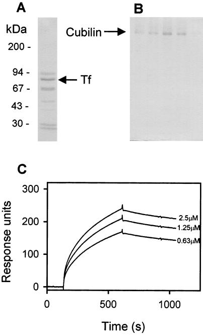
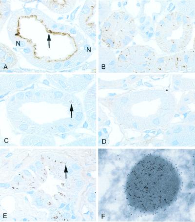
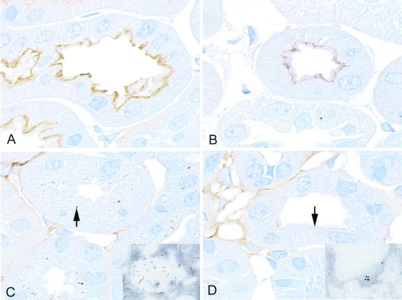
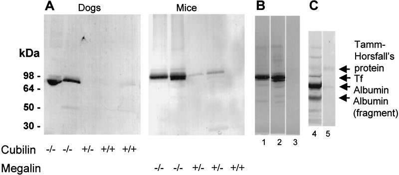
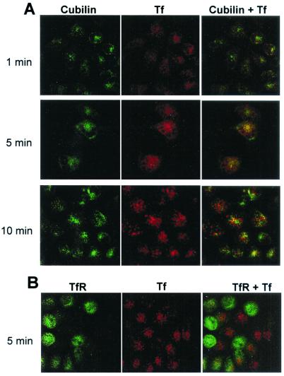
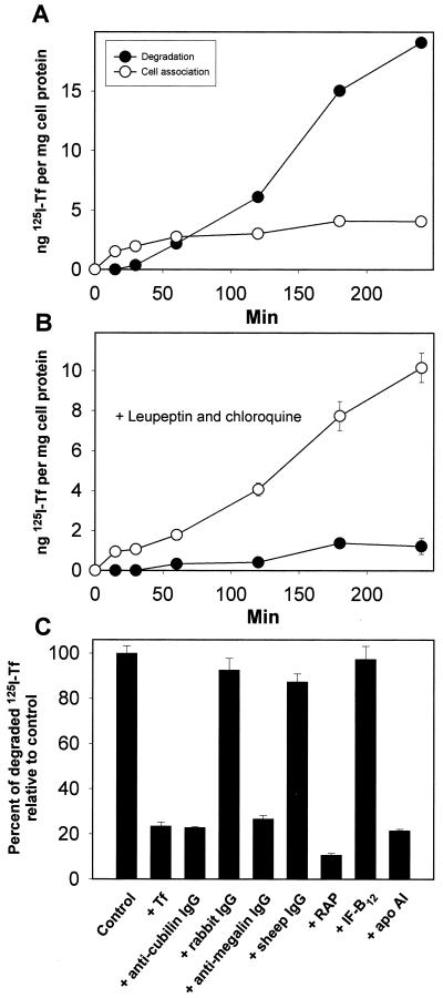
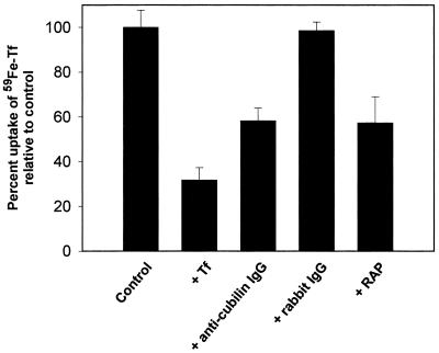
Cubilin is a 460-kDa protein functioning as an endocytic receptor for intrinsic factor vitamin B(12) complex in the intestine and as a receptor for apolipoprotein A1 and albumin reabsorption in the kidney proximal tubules and the yolk sac. In the present study, we report the identification of cubilin as a novel transferrin (Tf) receptor involved in catabolism of Tf. Consistent with a cubilin-mediated endocytosis of Tf in the kidney, lysosomes of human, dog, and mouse renal proximal tubules strongly accumulate Tf, whereas no Tf is detectable in the endocytic apparatus of the renal tubule epithelium of dogs with deficient surface expression of cubilin. As a consequence, these dogs excrete increased amounts of Tf in the urine. Mice with deficient synthesis of megalin, the putative coreceptor colocalizing with cubilin, also excrete high amounts of Tf and fail to internalize Tf in their proximal tubules. However, in contrast to the dogs with the defective cubilin expression, the megalin-deficient mice accumulate Tf on the luminal cubilin-expressing surface of the proximal tubule epithelium. This observation indicates that megalin deficiency causes failure in internalization of the cubilin-ligand complex. The megalin-dependent, cubilin-mediated endocytosis of Tf and the potential of the receptors thereby to facilitate iron uptake were further confirmed by analyzing the uptake of (125)I- and (59)Fe-labeled Tf in cultured yolk sac cells.


Figures

Figure 1
Figure 1
Identification of Tf as a cubilin ligand by affinity chromatography and surface plasmon resonance analysis. (A) SDS/PAGE and Coomassie staining of eluate from a cubilin-Sepharose column loaded with human serum. The band of Tf (identified by amino-terminal sequencing) is indicated. (B) SDS/PAGE and Coomassie staining of the eluate from a Tf-Sepharose column loaded with solubilized rat renal brush-border membranes. The band of cubilin (identified by immunoblotting) is indicated. (C) Surface plasmon resonance analysis of the binding of various concentrations of purified human Tf to immobilized cubilin.
Figure 2
Figure 2
Distribution of cubilin and Tf in normal dog kidney, in dog kidney with deficient cubilin surface expression, and in normal human kidney as determined by immunohistochemistry. (A) Cubilin staining is largely seen in the apical region (arrow) of the proximal tubule of normal dogs. Two nuclei (N) are marked. (B) A granular-like intracellular staining pattern is seen in dogs with deficient surface expression of cubilin. (C) A punctate staining (arrow) for Tf is seen in the proximal tubule of control dogs. (D) No Tf labeling is detectable in the dogs with affected cubilin expression. (E) Intensive accumulation of punctate Tf staining (arrow) in the human proximal tubule. (F) Immunoelectron microscopy identifies the human Tf-labeled structures as typical lysosomes. Immunoperoxidase staining, ×1,000 (A and E) and ×900 (B–D); immunogold staining, ×35,000 (F).
Figure 3
Figure 3
Distribution of cubilin and Tf in kidneys from wild-type and megalin-deficient mice as determined by immunohistochemistry. (A) Staining for cubilin in the apical part of the proximal tubule in the wild-type kidney. (B) Expression of cubilin in the megalin-deficient kidney. (C) Tf staining is seen as a vacuolar staining (arrow) in the proximal tubule of the wild-type kidney. (D) The megalin-deficient kidney exhibits an apical plasma membrane staining (arrow) for Tf corresponding to the surface staining for cubilin (B). (Insets) Immunogold staining for Tf in the wild-type (C) and the megalin-deficient (D) kidney.
Figure 4
Figure 4
Detection of Tf in urine of dogs with deficient surface expression of cubilin, in mice with deficient megalin expression, and in patients with Imerslund–Gräsbeck's syndrome. (A) Western blotting (nonreducing conditions) of 20-μl urine samples shows a high urinary content of Tf in animals homozygous (−/−) for cubilin or megalin deficiency, but not in heterozygous animals or control animals. (B) Western blotting for Tf in urine (2 μl) of two French children with Imerslund–Gräsbeck's disease (lanes 1 and 2) and in a control individual (lane 3). (C) Coomassie staining of 2 μl of urine from one of the Imerslund–Gräsbeck's disease patients with proteinuria (lane 4) and a control (lane 5). The filtered proteins and cubilin ligands, albumin and transferrin, are abundant proteins in the patient urine but not in normal urine. Also, several low molecular weight proteins are seen in the patient urine. The distale tubule protein, Tamm Horsfall's protein, is present in similar concentrations in patient and control urine.
Figure 5
Figure 5
Uptake of fluorescent Alexa 594-Tf by cultured rat yolk sac cells. (A) Uptake at 37°C of prebound Alexa 594-Tf (red) for 1, 5, or 10 min and immunostaining for cubilin using an Alexa 488-conjugated antibody (green). Note the initial colocalization of Tf and cubilin (1–5 min) and the subsequent vesicular accumulation of Tf in cubilin-negative structures (10 min). (B) Uptake at 37°C of prebound Alexa 594-Tf (red) for 5 min and immunostaining for TfR using an Alexa 488-conjugated antibody (green). Note the vesicular accumulation of Tf in TfR-negative cells.
Figure 6
Figure 6
Uptake and degradation of 125I-Tf by cultured rat yolk sac cells. Time course for cell association and degradation of 125I-Tf in the absence (A) or presence (B) of 100 μM leupeptin and 100 μM chloroquine. Degradation is measured as trichloroacetic acid-soluble 125I-labeled products released in the medium. (C) Degradation of 125I-Tf was assessed after 2 h at 37°C, in the presence of human Tf (1 mg/ml), human apolipoprotein (apo) A1 (1 μM), porcine IF-B12 (1 μM), RAP (1 μM), anti-cubilin polyclonal antibody (200 μg/ml), anti-megalin polyclonal antibody (200 μg/ml), and rabbit and sheep nonimmune IgG (200 μg/ml). Data represent percent of control values (incubation with buffer alone) and are means of triplicate determinations. Standard deviations are indicated where they exceed the size of the symbols.
Figure 7
Figure 7
Uptake of 59Fe in immortalized rat yolk sac cells incubated with 59Fe-Tf (1500 cpm/well). Inhibition of uptake by human Tf (1 mg/ml), anti-cubilin polyclonal antibody (200 μg/ml), rabbit IgG (200 μg/ml), and RAP (1 μM). The values are the mean ± 1 standard deviation of triplicate measurements.

References

    1. de Silva D M, Askwith C C, Kaplan J. Physiol Rev. 1996;76:31–47. - PubMed
    1. Fleming R E, Migas M C, Holden C C, Waheed A, Britton R S, Tomatsu S, Bacon B R, Sly W S. Proc Natl Acad Sci USA. 2000;97:2214–2219. . (First Published February 18, 2000; 10.1073/pnas.040548097) - PMC - PubMed
    1. Kawabata H, Yang R, Hirama T, Vuong P T, Kawano S, Gombart A F, Koeffler H P. J Biol Chem. 1999;274:20826–20832. - PubMed
    1. Kristiansen M, Graversen J H, Jacobsen C, Sonne O, Hoffman H J, Law S K, Moestrup S K. Nature (London) 2001;409:198–201. - PubMed
    1. Tolosano E, Hirsch E, Patrucco E, Camaschella C, Navone R, Silengo L, Altruda F. Blood. 1999;94:3906–3914. - PubMed

Publication types

Substances