. 2001 Oct 23;98(22):12630-5.
doi: 10.1073/pnas.221466798. Epub 2001 Oct 16.

Gambicin: a novel immune responsive antimicrobial peptide from the malaria vector Anopheles gambiae

Affiliations

Gambicin: a novel immune responsive antimicrobial peptide from the malaria vector Anopheles gambiae

J Vizioli et al. Proc Natl Acad Sci U S A. .

Abstract

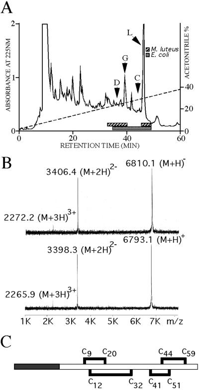
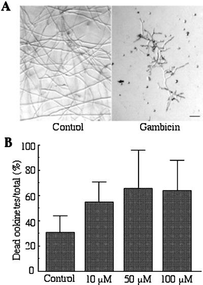
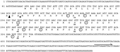
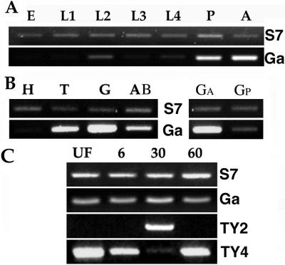
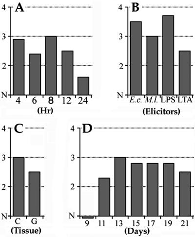
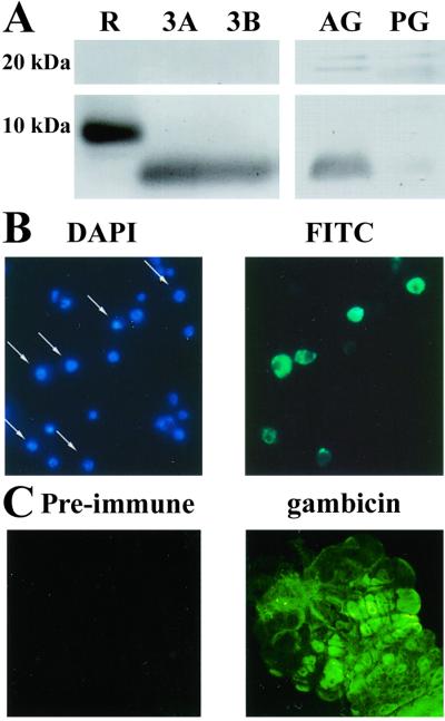
A novel mosquito antimicrobial peptide, gambicin, and the corresponding gene were isolated in parallel through differential display-PCR, an expressed sequence tag (EST) project, and characterization of an antimicrobial activity in a mosquito cell line by reverse-phase chromatography. The 616-bp gambicin ORF encodes an 81-residue protein that is processed and secreted as a 61-aa mature peptide containing eight cysteines engaged in four disulfide bridges. Gambicin lacks sequence homology with other known proteins. Like other Anopheles gambiae antimicrobial peptide genes, gambicin is induced by natural or experimental infection in the midgut, fatbody, and hemocyte-like cell lines. Within the midgut, gambicin is predominantly expressed in the anterior part. Both local and systemic gambicin expression is induced during early and late stages of natural malaria infection. In vitro experiments showed that the 6.8-kDa mature peptide can kill both Gram-positive and Gram-negative bacteria, has a morphogenic effect on a filamentous fungus, and is marginally lethal to Plasmodium berghei ookinetes. An oxidized form of gambicin isolated from the cell line medium was more active against bacteria than the nonoxidized form from the same medium.


Figures

Figure 1
Figure 1
(A) RP-HPLC of A. gambiae 4a-3B cell line conditioned medium. Fractions exhibiting antimicrobial activity against M. luteus and E. coli are indicated. Arrows indicate the fractions containing defensin (D), cecropin (C), gambicin (G), and lysozyme (L). (B) MALDI-TOF MS analysis of the purified oxidized (Upper) and nonoxidized (Lower) gambicins. Gambicin molecular mass values are expressed in m/z. Supplementary peaks corresponding to double- and triple-charged gambicin are indicated. (C) Primary structure of the 61-aa mature gambicin disulfide bridge array. The cysteine pairing is indicated in bold lines. The20-residue signal peptide–propeptide region is shown in black.
Figure 2
Figure 2
(A) Antifungal assay. Light microscopy observation of N. crassa hyphal growth after 24 h of incubation with water (Control) or 100 μM gambicin. (Scale bar, 100 μm.) (B) Antiparasitic assay. Percentage of dead ookinetes after incubation with PBS (Control) or gambicin, tested at different concentrations.
Figure 3
Figure 3
Nucleotide and encoded amino acid sequence of the gambicin cDNA. The priming site for the random 10-mer primer used in the differential display amplification of the 3′ UTR gambicin fragment is indicated by a horizontal arrow. The predicted signal peptide cleavage site according to von Heine and the beginning of the mature peptide are marked with an arrowhead and asterisk, respectively. Circles mark the 8 cysteines.
Figure 4
Figure 4
RT-PCR expression analysis of gambicin mRNA. cDNA templates were normalized by using a ribosomal protein S7-specific primer pair. (A) Developmental expression profile assayed in embryo (E), four larval stages (L1–L4), pupae (P), and adult females (A). (B) Gambicin expression in head (H), thorax (T), midgut (G), abdomen (AB), anterior midgut (GA), and posterior midgut (GP) of 4-day-old female adult mosquitoes. (C) Expression of gambicin in unfed (UF) and blood-fed adult female mosquitoes at 6, 30, and 60 h after feeding. Expression of the blood feeding regulated digestive trypsins, Antryp 2 (TY2), and Antryp 4 (TY4) were also assayed in the same samples as controls.
Figure 5
Figure 5
Infection inducibility of gambicin transcription in bacteria-challenged cell lines and malaria-challenged mosquitoes. The RT-PCR products were analyzed on Cybr-Green-stained agarose gels and quantified by using a phosphorimager. The templates were normalized for expression of the ribosomal protein S7 gene and the relative expression levels for gambicin were arithmetically adjusted for equal S7 expression in the compared templates. Experiments were repeated three to four times and the average fold regulation was estimated for three experiments and indicated in the figure. Variations between experiments ranged between 0.1- and 0.6-fold regulation. The y axes indicate fold regulation below or above the naive (N) expression level. Transcriptional induction of gambicin in: (A) the 4a-3B cell line at 4, 6, 8, 12, and 24 h after challenged with a mixture of E. coli and M. luteus; (B) the 4a-3B cell line at 6 h after challenge with E. coli, M. luteus, LPS, and LTA; (C) malaria-infected mosquito carcass (C) and midgut (G) at 24 h after feeding on a P. berghei-infected mouse; (D) malaria-infected mosquitoes at 9, 11, 13, 15, 17, 19, and 21 days after feeding on a P. berghei-infected mouse.
Figure 6
Figure 6
(A) Immunoblot analysis of gambicin protein content in anterior (AG) and posterior (PG) midgut tissue extracts and 4a-3A (3A) and 4a-3B (3B) cell line conditioned medium. Recombinant gambicin (R) was used as a control. (B) 4′,6-diamidino-2-phenylindole (DAPI) and FITC (gambicin immunofluorescence) staining of 4a-3B cells. A subset of the nuclear DAPI-stained cells exhibit strong gambicin protein content. (C) Whole-mount antibody staining of anterior midgut giving high signals for gambicin in cells of the cardia. Control panel shows midgut staining with rabbit preimmune serum.

References

    1. Beier J C. Annu Rev Entomol. 1998;43:519–543. - PubMed
    1. Collins F H, Sakai R K, Vernick K D, Paskewitz S, Seeley D C, Miller L H, Collins W E, Campbell C C, Gwadz R W. Science. 1986;234:607–610. - PubMed
    1. Vernick K D, Fujioka H, Seeley D C, Tandler B, Aikawa M, Miller L H. Exp Parasitol. 1995;80:583–595. - PubMed
    1. Ghosh A, Edwards M J, Jacobs-Lorena M. Parasitol Today. 2000;16:196–201. - PubMed
    1. Dimopoulos G, Seeley D, Wolf A, Kafatos F C. EMBO J. 1998;17:6115–6123. - PMC - PubMed

Publication types

Associated data