. 2001 Nov 6;98(23):13108-13.
doi: 10.1073/pnas.231148598. Epub 2001 Oct 23.

Calcineurin controls nerve activity-dependent specification of slow skeletal muscle fibers but not muscle growth

Affiliations

Calcineurin controls nerve activity-dependent specification of slow skeletal muscle fibers but not muscle growth

A L Serrano et al. Proc Natl Acad Sci U S A. .

Abstract

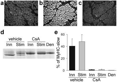
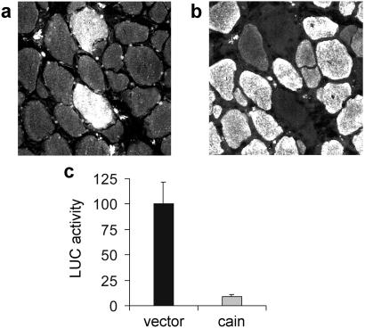
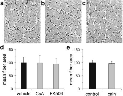
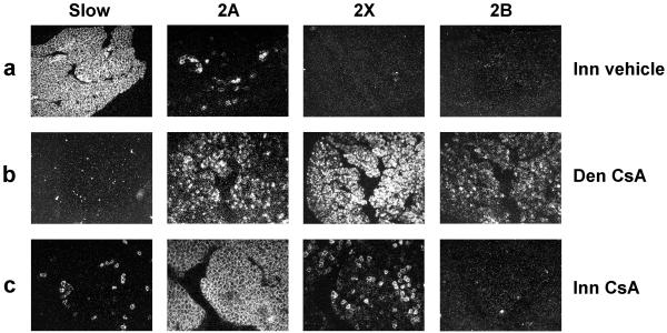
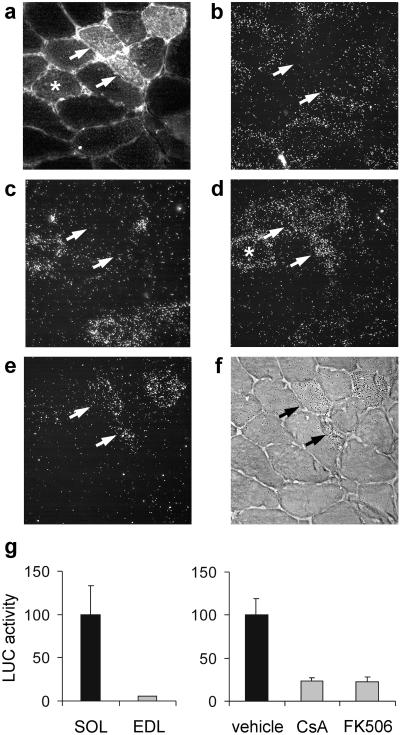
Nerve activity can induce long-lasting, transcription-dependent changes in skeletal muscle fibers and thus affect muscle growth and fiber-type specificity. Calcineurin signaling has been implicated in the transcriptional regulation of slow muscle fiber genes in culture, but the functional role of calcineurin in vivo has not been unambiguously demonstrated. Here, we report that the up-regulation of slow myosin heavy chain (MyHC) and a MyHC-slow promoter induced by slow motor neurons in regenerating rat soleus muscle is prevented by the calcineurin inhibitors cyclosporin A (CsA), FK506, and the calcineurin inhibitory protein domain from cain/cabin-1. In contrast, calcineurin inhibitors do not block the increase in fiber size induced by nerve activity in regenerating muscle. The activation of MyHC-slow induced by direct electrostimulation of denervated regenerating muscle with a continuous low frequency impulse pattern is blocked by CsA, showing that calcineurin function in muscle fibers and not in motor neurons is responsible for nerve-dependent specification of slow muscle fibers. Calcineurin is also involved in the maintenance of the slow muscle fiber gene program because in the adult soleus muscle, cain causes a switch from MyHC-slow to fast-type MyHC-2X and MyHC-2B gene expression, and the activity of the MyHC-slow promoter is inhibited by CsA and FK506.


Figures

Figure 1
Figure 1
CsA and FK506 block the expression of MyHC-slow in regenerating soleus muscle. (a–d) Immunofluorescence analysis of sections stained with an antibody specific for MyHC-slow. Note that regenerating soleus muscles from rats treated with CsA (b) or FK506 (c) show a decreased proportion of reactive fibers compared with rats treated with vehicle (a). The percentage of fibers expressing MyHC-slow in the three experimental groups is shown in d. (e) SDS/PAGE profile of MyHCs (Upper). Note that the high mobility MyHC-slow band (arrow) is abundant in control muscle (left lane) but is barely detectable after treatment with CsA (center lane) or FK506 (right lane). This decrease is confirmed by quantitation of densitometric values of the percentage of MyHC-slow relative to all MyHCs (Lower). (f) MyHC-slow promoter activity is down-regulated by CsA and FK506. A plasmid containing the MyHC-slow promoter linked to a luciferase reporter gene was injected in regenerating muscles, and luciferase activity was measured 7 days later in tissue homogenates. Luciferase (LUC) activity after treatment with calcineurin inhibitors is expressed as the percentage of that measured in rats treated with vehicle.
Figure 2
Figure 2
CsA prevents the up-regulation of MyHC-slow induced by electrostimulation in denervated regenerating soleus muscle. Muscles were denervated and electrostimulated with a tonic 20-Hz pattern of impulses that resembles the firing pattern of slow motor neurons. (a–c) Immunofluorescence analysis with anti-MyHC-slow antibody of regenerating soleus muscles after denervation (a), denervation and electrostimulation (b), or denervation and electrostimulation plus treatment with CsA (c). Note that the up-regulation of MyHC-slow induced by electrostimulation is prevented by CsA. (d and e) Electrophoretic analysis of MyHCs shows that the high mobility MyHC-slow band is abundant in muscles innervated (Inn) or denervated and electrostimulated (Stim) but is undetectable in denervated muscles (Den). CsA treatment blocks the up-regulation of MyHC-slow in both innervated and electrostimulated muscles.
Figure 3
Figure 3
The calcineurin inhibitor cain/cabin-1 prevents the up-regulation of MyHC-slow and the activation of MyHC-slow promoter in regenerating soleus muscle. Serial cross sections of regenerating soleus muscle transfected with myc-tagged cain and stained with anti-myc (a) or anti-MyHC-slow (b) antibodies. Note that the two fibers expressing myc-tagged cain do not express MyHC-slow, unlike most surrounding untransfected fibers. (c) Cotransfection of MyHC-slow promoter and cain shows that MyHC-slow promoter activity is down-regulated by cain.
Figure 4
Figure 4
Muscle fiber growth in regenerating muscle is not affected by calcineurin inhibitors. (a–c) Hematoxylin and eosin staining of cross sections of soleus muscle from control (a), CsA-treated (b), and FK506-treated (c) animals shows normal growth of regenerating muscle fibers at day 10 after injury. (d) Fiber size is unchanged in CsA- and FK506-treated compared with vehicle-treated animals. (e) Fiber size is unchanged in fibers transfected with myc-tagged cain compared with untransfected surrounding fibers (control).
Figure 5
Figure 5
CsA blocks the MyHC-2A → MyHC-slow switch but not the MyHC-2X (-2B) → MyHC-2A switch in regenerating soleus muscle. Serial cross sections of innervated regenerating soleus muscle were processed for in situ hybridization with probes specific for MyHC-slow, MyHC-2A, MyHC-2X, and MyHC-2B transcripts and viewed by dark-field microscopy. Note that innervated muscles (Top) show a predominance of MyHC-slow transcripts with a minor population of fibers expressing MyHC-2A transcripts. In contrast, denervated muscles both in rats treated with vehicle (not shown) or CsA (Middle) show a predominance of MyHC-2X and MyHC-2B with minor amounts of MyHC-2A but complete absence of MyHC-slow. CsA treatment (Bottom) blocks almost completely the switch to MyHC-slow in innervated muscles, whereas the transition from MyHC-2X (-2B) to MyHC-2A is almost unaffected, thus leading to a predominant expression of MyHC-2A transcripts with a minor population of fibers expressing MyHC-2X transcripts.
Figure 6
Figure 6
MyHC-slow gene expression is down-regulated by calcineurin inhibitors in the adult soleus muscle. (a–f) Serial cross sections of adult soleus transfected with myc-cain were processed for immunofluorescence with anti-myc antibody (a) or for in situ hybridization with probes specific for MyHC-slow (b), MyHC-2A (c), MyHC-2X (d), and MyHC-2B transcripts (e and f) and viewed by dark-field microscopy (b–e). MyHC-2B transcript distribution is also illustrated by light-field microscopy (f) to allow direct comparison with myc-cain distribution. Note that fibers strongly reactive for myc-cain (arrows) contain MyHC-2X and MyHC-2B but not MyHC-2A and MyHC-slow transcripts, whereas fibers weakly reactive for myc-cain (asterisk) express MyHC-2X but not MyHC-2B. (g) MyHC-slow promoter activity is higher in the adult slow soleus (SOL) compared with the fast extensor digitorum longus (EDL) muscle (Left) and is markedly inhibited by CsA and FK506 in soleus muscle (Right).

References

    1. Schiaffino S, Reggiani C. Physiol Rev. 1996;76:371–423. - PubMed
    1. DiMario J X, Stockdale F E. Dev Biol. 1997;188:167–180. - PubMed
    1. Hughes S M, Salinas P C. Curr Opin Neurobiol. 1999;9:54–64. - PubMed
    1. Pette D, Vrbova G. Rev Physiol Biochem Pharmacol. 1992;120:115–202. - PubMed
    1. Chin E R, Olson E N, Richardson J A, Yang Q, Humphries C, Shelton J M, Wu H, Zhu W, Bassel-Duby R, Williams R S. Genes Dev. 1998;12:2499–2509. - PMC - PubMed

Publication types

LinkOut - more resources