. 2001 Nov 6;98(23):13055-9.
doi: 10.1073/pnas.231364798. Epub 2001 Oct 23.

Reactivation of proliferin gene expression is associated with increased angiogenesis in a cell culture model of fibrosarcoma tumor progression

Affiliations

Reactivation of proliferin gene expression is associated with increased angiogenesis in a cell culture model of fibrosarcoma tumor progression

D J Toft et al. Proc Natl Acad Sci U S A. .

Abstract

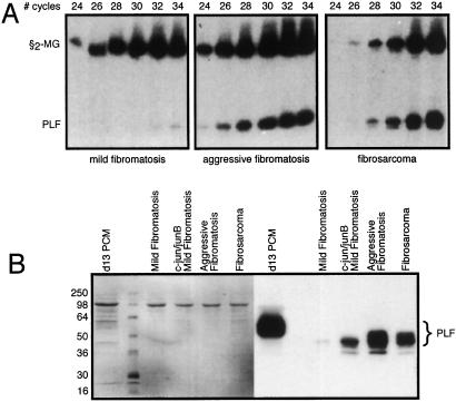
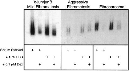
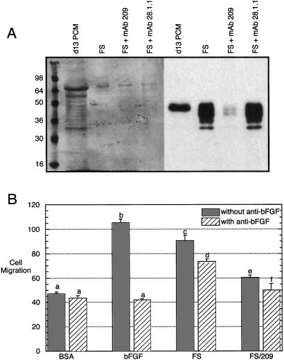
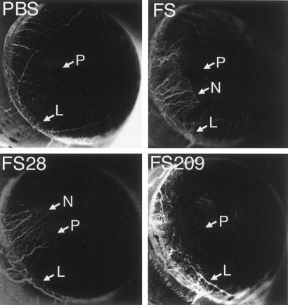
Proliferin (PLF) is an angiogenic placental hormone. We now report that PLF gene expression can also occur in a progressive fibrosarcoma mouse tumor cell model. PLF mRNA and protein are detectable at very low levels in cell lines derived from the mild noninvasive stage of tumor development. Expression is greatly augmented in cell lines from the aggressively invasive stage of development, a stage at which the tumor becomes highly angiogenic, and PLF expression remains high in cell lines from the end stage of fibrosarcoma. Activator protein 1 factors present at high levels in the more invasive stages of the tumor may in part allow for increased PLF expression, as cells from the mild stage in which c-jun and junB are stably expressed secrete levels of PLF comparable to that of the advanced stages. Secreted PLF protein is functionally important in tumor cell angiogenic activity, as demonstrated by the reduction of angiogenic activity in fibrosarcoma cell culture medium by immunodepletion of PLF. These results suggest that an extraembryonic genetic program, which has evolved to support fetal growth, may be reactivated in certain tumors and contribute to tumor growth.


Figures

Figure 1
Figure 1
Tumor stage-specific expression of PLF. (A) RT-PCR with primers specific for PLF and β2-macroglobulin mRNAs was carried out with total RNA from mild fibromatosis (Left), aggressive fibromatosis (Center), and fibrosarcoma (Right) cells. Products were analyzed after 24–34 PCR cycles. The amount of PLF mRNA detected at the fibrosarcoma stage is ≈20-fold greater than the level at the mild fibromatosis stage. (B) Serum-free media were collected from cultured mild fibromatosis, aggressive fibromatosis, and fibrosarcoma cells, and from mild fibromatosis cells stably expressing the AP-1 factors c-jun and junB. Proteins in these samples were fractionated by gel electrophoresis and PLF then detected by immunoblotting (Right). Medium from cultured placental tissue collected on day 13 of gestation served as a positive control, and Ponceau-S staining (Left) demonstrates equal protein loading.
Figure 2
Figure 2
Serum and glucocorticoid regulation of PLF expression in tumor cells. Mild fibromatosis cells expressing c-jun and junB, aggressive fibromatosis cells, and fibrosarcoma cells were serum-starved for 48 h in the absence or presence of 0.1 μM dexamethasone or starved and then stimulated with 15% FBS for 24 h, again with or without dexamethasone. Total RNA from these cells was fractionated by formaldehyde gel electrophoresis, transferred to a filter, and hybridized to detect PLF mRNA.
Figure 3
Figure 3
PLF1 expression in fibrosarcoma cells. (A) Schematic diagrams of the four known PLF mRNA sequences are shown with the locations of cleavage sites for the BstXI, SatI, and NdeI restriction endonucleases. Fragment sizes in base pairs that would result from cleavage with these enzymes are listed below. (B) PLF1, PLF2, and fibrosarcoma cDNAs were amplified by PCR, using primers corresponding to nucleotide sequences found in all PLF transcripts. Reaction products were digested with the indicated restriction endonucleases, resolved by PAGE, and detected by ethidium bromide staining. Size markers are labeled on the left; all expected fragment sizes are listed on the right.
Figure 4
Figure 4
Cell culture angiogenic activity secreted by fibrosarcoma cells . (A) Medium collected from cultured fibrosarcoma cells (FS) was untreated or was incubated with mAb 209 (specific for PLF) or 28.1.1 (nonspecific control). Ab-antigen complexes were removed by using protein G beads. After these treatments, the medium was analyzed by gel electrophoresis and immunoblotting for the removal of PLF protein. Medium from cultured mouse placentas isolated on day 13 of gestation was used as a positive control for PLF detection; Ponceau-S staining shows equal protein loading (Left). (B) Basal endothelial cell migration was measured in a Boyden chamber in the presence of 0.1% BSA, and maximally induced migration was determined after addition of 10 ng/ml of bFGF. Fibrosarcoma culture medium, either intact or after removal of PLF, was also tested. Each sample was also assayed after addition of a neutralizing Ab against bFGF. Results were analyzed by a one-way ANOVA followed by a Tukey's post hoc test. n = 4, and the results were reproduced with an independently generated set of samples. a vs. b, c, or d: P < 0.001; c vs. d: P < 0.01; c vs. e: P < 0.001; d vs. f: P < 0.001.
Figure 5
Figure 5
In vivo angiogenic activity secreted by fibrosarcoma cells. Hydron pellets were prepared containing buffer (PBS), fibrosarcoma culture-conditioned medium (FS), tumor cell medium treated with anti-PLF-related protein mAb 28.1.1, which does not recognize PLF (FS28), or tumor cell medium from which PLF had been removed by immunodepletion with mAb 209 (FS209). The pellets were placed in rat corneas, and neovascularization was monitored after 7 days. Vigorous ingrowth of the new vessels from the limbus (L) in the direction of the pellet (P) was scored as neovascularization (N). Inverted grayscale images are shown to enhance visualization of neovascularization. Positive responses were detected in 0/4 corneas with PBS, 4/4 with FS, 3/4 with FS28, and 0/4 for FS209.

References

    1. Soares M J, Muller H, Orwig K E, Peters T J, Dai G. Biol Reprod. 1998;58:273–284. - PubMed
    1. Galosy S S, Talamantes F. Endocrinology. 1995;136:3993–4003. - PubMed
    1. Müller H, Liu B, Croy B A, Head J R, Hunt J S, Dai G, Soares M J. Endocrinology. 1999;140:2711–2720. - PubMed
    1. Lin J, Linzer D I H. J Biol Chem. 1999;274:21485–21489. - PubMed
    1. Nilsen-Hamilton M, Hamilton R T, Alvarez-Azaustre E. Gene. 1987;51:163–170. - PubMed

Publication types

MeSH terms