. 2001 Nov 6;98(23):13312-7.
doi: 10.1073/pnas.241338598. Epub 2001 Oct 23.

Invasive melanoma in Cdk4-targeted mice

Affiliations

Invasive melanoma in Cdk4-targeted mice

R Sotillo et al. Proc Natl Acad Sci U S A. .

Abstract

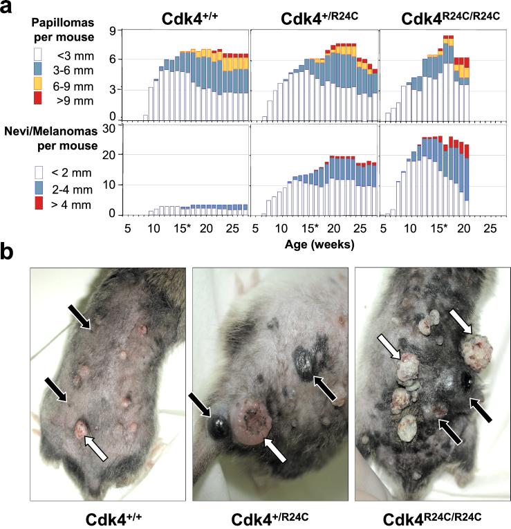
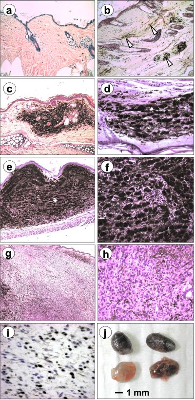
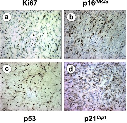
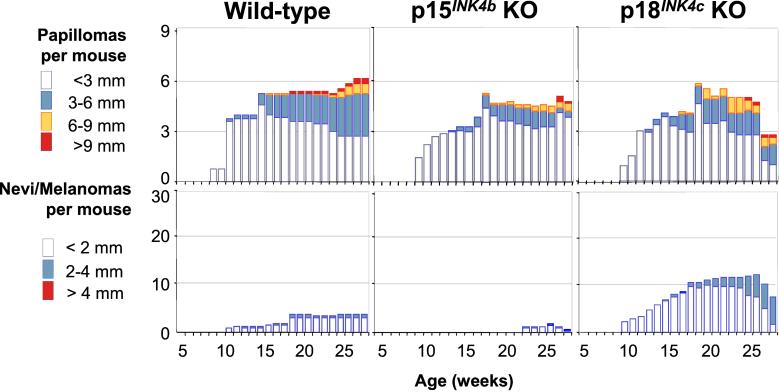
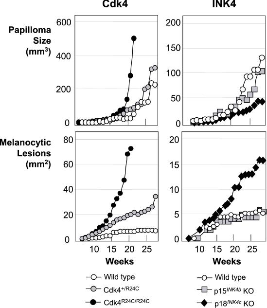
Many human tumors harbor mutations that result in deregulation of Cdk4 activity. Most of these mutations involve overexpression of D-type cyclins and inactivation of INK4 inhibitors. In addition, a mutation in the Cdk4 protein has been described in patients with familial melanoma (Wolfel, T., Hauer, M., Schneider, J., Serrano, M., Wolfel, C., et al. (1995) Science 269, 1281-1284; Zuo, L., Weger, J., Yang, Q., Goldstein, A. M., Tucker, M. A., et al. (1996) Nat. Genet. 12, 97-99). This mutation, R24C, renders the Cdk4 protein insensitive to inhibition by INK4 proteins including p16(INK4a), a major candidate for the melanoma susceptibility locus. Here we show that knock-in mice expressing a Cdk4 R24C allele are highly susceptible to melanoma development after specific carcinogenic treatments. These tumors do not have mutations in the p19(ARF)/p53 pathway, suggesting a specific involvement of the p16(INK4a)/Cdk4/Rb pathway in melanoma development. Moreover, by using targeted mice deficient for other INK4 inhibitors, we show that deletion of p18(INK4c) but not of p15(INK4b) confers proliferative advantage to melanocytic tumor growth. These results provide an experimental scenario to study the role of Cdk4 regulation in melanoma and to develop novel therapeutic approaches to control melanoma progression.


Figures

Figure 1
Figure 1
Development of skin lesions in mice treated with DMBA + TPA. (a) The average number of papillomas (top panels) or macroscopic nevi/melanomas (lower panels) per mouse was scored, grouped by size (diameter of the lesions in mm), and represented for each genotype: Cdk4+/+ (n =13), Cdk4+/R24C (n =13), and Cdk4R24C/R24C (n =20). Because a significant fraction of Cdk4R24C/R24C mice are dead by week 21, the statistical representation of their skin lesions was stopped at week 20. (b) Representative pictures taken at week 20. White arrows indicate papillomas, and black arrows show the position of small nevi (Cdk4+/+) or melanomas (Cdk4+/R24C and Cdk4R24C/R24C).
Figure 2
Figure 2
Representative sections of nevi and melanomas in Cdk4 R24C mice. Hematoxylin and eosin images of Cdk4+/+ (a) and Cdk4R24C/R24C (b) mice after treatment with the DMBA + TPA protocol are shown. At this magnification (×200), melanocytes are barely visible in wild-type mice but are abundant in Cdk4R24C/R24C (arrow heads). (c and d) Small benign lesions similar to human blue nevus. (e and f) Cellular lesions with compacted groups of spindle-shaped melanocytes with a small load of melanin. (g and h) Invasive melanomas with evident loss of melanin content and infiltration to nearby tissues. (i) Melanocytic origin of advanced lesions was determined by positive immunostaining for the S100 antigen. Original magnifications: a and c, ×100; b, d, and f, ×400; e, ×45; g, ×630. (j) Enlarged lymph nodes with high melanin content commonly found in Cdk4R24C/R24C mice with primary melanomas.
Figure 3
Figure 3
Representative staining of Cdk4R24C/R24C melanomas against Ki67 proliferative antigen (a), p16INK4a (b), p53 (c), and p21Cip1 (d). Original magnification is ×400.
Figure 4
Figure 4
Development of papillomas and melanocytic lesions in INK4-deficient mice. The average number of skin lesions per mouse is represented, and the same conventions described for Fig. 1 are maintained. The following genotypes were analyzed: wild type (n = 9), p15INK4b null (p15INK4b KO, n = 8), and p18INK4c null (p18INK4c KO, n =15). INK4a-ARF null mice (n =14) were treated also, but these animals died before 12 weeks without obvious skin lesions and are not represented in the figure.
Figure 5
Figure 5
Growth kinetics of papillomas or melanocytic lesions in Cdk4- (Left) or INK4- (Right) targeted mice. Tumor volume (mm3) or area (mm2) was scored and represented against time, because the lesion appears in the skin (0 weeks). Because melanocytic lesions are flat in the early stages, the area is represented, and the vertical phase of growth therefore is not considered. Please note that the size of lesions is represented at a different scale for Cdk4- and INK4-targeted mice.

References

    1. Sherr C J. Cancer Res. 2000;60:3689–3695. - PubMed
    1. Hirama T, Koeffler H P. Blood. 1995;86:841–854. - PubMed
    1. Hall M, Peters G. Adv Cancer Res. 1996;68:67–108. - PubMed
    1. Ruas M, Peters G. Biochim Biophys Acta. 1998;1378:F115–F177. - PubMed
    1. Roussel M F. Oncogene. 1999;18:5311–5317. - PubMed

Publication types

MeSH terms

Substances