

Hepatic trisegmentectomy and other liver resections
- PMID: 1162576
- PMCID: PMC2653783
Hepatic trisegmentectomy and other liver resections
Abstract
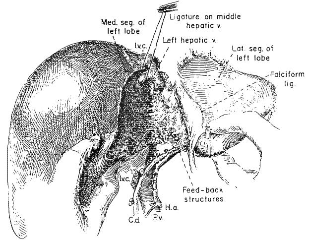
Trisegmentectomy, extended right hepatic lobectomy, is the removal of the true right lobe of the liver in continuity with most or all of the medial segment of the left lobe. Some important features of the operation have not been well described previously. To perform trisegmentectomy safely, a fusion of liver tissue covering the umbilical fissure at the level of the falciform ligament must first be split open in many patients. The left branches of the portal triad structures are mobilized from the undersurface of the liver nearly to but not into the umbilical fissure. The blood supply and duct drainage of the medial segment originate within the umbilical fissure and feed back toward the right side buried in liver substance. They are found with blunt dissection just to the right of the falciform ligament, encircled and ligated. Failure to appreciate this switch back anatomic arrangement may lead to injury of the blood supply or biliary drainage of the residual lateral segment. Parenthetically, the mirror image operation of lateral segmentectomy could result in devascularization of the medial segment if dissection and ligation were performed within the umbilical fissure instead of well to the left of this landmark. In most trisegmentectomies, the left portion of the caudate lobe is not removed. This small piece of tissue is interposed between the lateral segment and the inferior vena cave into which it drains by small tributaries. If the left portion of the caudate lobe is to be excised, it is necessary to ligate the last two posteriorly running branches before the main left trunks of the portal triad structures reach the umbilical fissure. Once this step is taken and if the caudate removal is completed, the remaining lateral segment usually has only one remaining outflow, that of the left hepatic vein. The other principles of trisegmentectomy are the same as with less radical subtotal hepatic resection. These include vascular suture closure of the main outflow veins, avoidance of parasegmental planes that leave behind a strip of devitalized tissue, preservation of intersegmental or interlobar veins, omission of techniques that sew shut or otherwise cover the raw surface of the remnant and provision of adequate drainage of dead space. After trisegimentectomy and also after true lobectomy, this last objective is usually met by leaving part of the operative incision open. Using these guidelines, there has been no mortality with 27 hepatic resections carried out since 1963, including 14 trisegmentectomies.
Figures


References
-
- BRUNSCHWIG A. The surgery of hepatic neoplasms with special reference to right and left lobectomies. XVI Congress de la Societe Internationale de Chirurgie, Copenhagen. Imprimerie Medicale et Scientifique; Brussels: 1955. pp. 1122–1132.
-
- COUINAUD C. Le Foie. Etudes Anatomiques et Chirurgicales. Masson; Paris: 1957.
-
- GOLDSMITH NA, WOODBURNE RT. The surgical anatomy pertaining to liver resection. Surg. Gynecol. Obstet. 1957;105:310. - PubMed
-
- HEALEY JE., JR. Clinical anatomic aspects of radical hepatic surgery. J. Int. Coll. Surg. 1954;22:542. - PubMed
Publication types
MeSH terms
Grants and funding
LinkOut - more resources
Full Text Sources
Miscellaneous