. 2001 Oct;128(20):3899-912.
doi: 10.1242/dev.128.20.3899.

An isoform of eIF4E is a component of germ granules and is required for spermatogenesis in C. elegans

Affiliations

An isoform of eIF4E is a component of germ granules and is required for spermatogenesis in C. elegans

A Amiri et al. Development. 2001 Oct.

Abstract

Control of gene expression at the translational level is crucial for many developmental processes. The mRNA cap-binding protein, eIF4E, is a key player in regulation of translation initiation; appropriate levels of eIF4E are essential for normal cell-cycle regulation and tissue differentiation. The observation that eIF4E levels are elevated during gametogenesis in several organisms suggests that eIF4E might have a specific role in gamete formation as well. We show that one of the five isoforms of C. elegans eIF4E, IFE-1, is enriched in the germline and is a component of germ granules (P granules). The association of IFE-1 with P granules requires the P-granule protein PGL-1. In vitro PGL-1 interacts directly with IFE-1, but not with the other four isoforms of eIF4E. Analysis of animals depleted of IFE-1 by RNAi shows that IFE-1 is required for spermatogenesis, specifically for efficient progression through the meiotic divisions and for the production of functional sperm, in both hermaphrodites and males. The requirement for IFE-1 is highly sensitive to temperature. IFE-1 is not required for oogenesis, as ife-1(RNAi) hermaphrodites produce viable progeny when normal sperm are supplied. Consistent with a primary role in spermatogenesis, ife-1 mRNA levels are highest in regions of the gonad undergoing spermatogenesis. Our results suggest that C. elegans spermatogenesis requires either this specific isoform of eIF4E or an elevated level of eIF4E.


Figures

Fig. 1
Fig. 1
IFE-1 and PGL-1 proteins interact directly in vitro. [35S]IFE proteins were tested for binding to GST-PGL-1. Bound proteins were analyzed by SDS-PAGE on 12% gels. The input lanes contain 10% of the amount of radiolabeled protein used in the binding assays. The identity of the upper band in the IFE-1 input lane, which is also present in other IFE input lanes, is not known. (A) [35S]IFE proteins were tested for binding to GST-PGL-1 or GST as indicated. (B) [35S]IFE-1 was treated with RNase A or water, before incubating with GST-PGL-1. The slight variation in intensity of the [35S]IFE-1 band in the ‘+’ and ‘-’ lanes was not observed in a duplicate experiment. The Ethidium Bromide-stained agarose gel of RNA extracted after RNase A treatment or mock treatment shows that endogenous RNAs were reduced to below detection by RNase A treatment.
Fig. 2
Fig. 2
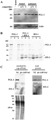
PGL-1 is retained by m7GTP-Sepharose through interaction with IFE-1. (A) Retention of native PGL-1 on m7GTP-Sepharose. Cleared lysates were prepared from wild-type (N2) worms and incubated with m7GTP-Sepharose. Bound proteins were eluted with m7GTP as described in Materials and Methods. To verify the specificity of cap interaction, either 100 μM GTP (G), 100 μM m7GTP (m7G) or buffer A (-) were added to lysates as competitors before chromatography. Eluted fractions (bound; ~0.3 μg protein), column flow-through (unbound, 10% volume; ~24 μg protein) and original lysate (lysate, 10% volume; ~30 μg protein) were resolved by SDS-PAGE on a 6% gel and immunoblotted using anti-PGL-1 antiserum or anti-chicken actin antiserum (Sigma). (B) Retention of [35S]PGL-1 on m7GTP-Sepharose via [35S]IFE-1. [35S]PGL-1 synthesized in reticulocyte lysate (total, t) is not retained on m7GTP-Sepharose (bound, b) unless IFE-1 is also synthesized in the lysate. Instead, PGL-1 alone is found entirely in the column flow-through (unbound, u). Equivalent volumes of total, bound and unbound fractions were resolved by SDS-PAGE on a 10% gel and 35S-labeled proteins visualized by a phosphorimager. The asterisk indicates the migration of a protein of unknown identify (as seen in Fig. 1). (C) Depletion of IFE-1 in ife-1(RNAi) worms (generated by feeding; see Materials and Methods). m7GTP-binding proteins were enriched from wild-type (N2) or ife-1(RNAi) worm extracts, and analyzed by western blot (1.4 μg protein per lane) in order to detect endogenous IFE-1 and cap-retained PGL-1. Silver staining of a similar gel verified equal loading of m7GTP-binding proteins. Western blotting of total protein (30 μg per lane) from worm extracts verified the presence of PGL-1 and another eIF4E isoform, IFE-2, in ife-1(RNAi) worms.
Fig. 3
Fig. 3
ife-1 mRNA is enriched in the germline. (A) Northern analysis of ife-1 mRNA. Poly(A)+ RNA was prepared from synchronous populations of wild-type (N2) and glp-4(bn2ts) hermaphrodites grown at 25°C, at which temperature glp-4 mutants produce a severely underproliferated germline. The rpp-1 mRNA, which encodes a ribosomal protein, was used as a loading control. Relative levels of ife-1 mRNA are shown at the bottom. (B) Distribution of ife-1 mRNA in embryos, as revealed by whole-mount in situ hybridization. The cDNA clone yk504h9 was used as a probe. ife-1 mRNA is present in all cells until the 100-200-cell stage and gradually disappears by the 300-400-cell stage. (C) Distribution of ife-1 mRNA in larvae and adult hermaphrodites. ife-1 mRNA first becomes detectable in the germline of L3 stage larvae and is maintained in the germline throughout adulthood. The signal is more intense in the regions of the gonad undergoing spermatogenesis (carets).
Fig. 4
Fig. 4
IFE-1, IFE-3 and IFE-5 are enriched in the germline. m7GTP affinity-purified proteins derived from 1.5 mg of total protein from wild-type (N2) or germline-deficient (glp-4) worms were resolved by SDS-PAGE on 6% (A) and 12% (B) gels. (A) The gel on the left was stained with silver nitrate, the gel on the right was immunoblotted using anti-PGL-1 antiserum. (B) The gels were immunoblotted using antibodies to isoform-specific C-terminal peptides for each of the IFE proteins. Both wild-type and glp-4 worms were grown at 25°C, the restrictive temperature for germline development in the glp-4 strain.
Fig. 5
Fig. 5
IFE-1∷GFP is associated with P granules in wild-type (N2) but not in pgl-1 embryos. The genotypes of embryos are listed above the columns. Embryos measure ~30×50 μm. (A,E) Fluorescence micrographs of 16-20-cell stage embryos. IFE-1∷GFP is localized in perinuclear granules in the germline cell (P4) in N2 but not in pgl-1. (H) Fluorescence micrograph of a four-cell stage embryo. IFE-1∷GFP signal is not detected in the cytoplasm or P granules of RNAi embryos. (B-D,F,G) Immunofluorescence micrographs of 70-90-cell stage embryos co-stained with anti-GFP (B,F) and anti-GLH-2 (C,G) antibodies. Anti-GFP staining is detectable in perinuclear granules in the germ cells (Z2 and Z3) of N2 but not in pgl-1 germ cells. (D) The merged image of B,C demonstrates double staining of P granules by anti-GFP and anti-GLH-2 antibodies in N2.
Fig. 6
Fig. 6
ife-1(RNAi) worms display temperature-sensitive sterility and reduced brood size. The F1 hermaphrodite progeny of mothers injected with ife-1 dsRNA or with water (control) were analyzed for sterility and brood size at (A) 16°C, (B) 20°C and (C) 25°C. The numbers of ife-1(RNAi) F1 worms analyzed were 14, 53 and 225 at 16°C, 20°C and 25°C, respectively. The numbers of control F1 progeny analyzed were 13, 13 and 14 at 16°C, 20°C and 25°C, respectively. At 25°C, 80% of RNAi worms did not produce any progeny. The remaining 20% of RNAi worms at 25°C and all RNAi worms at 16°C and 20°C produced a smaller number of progeny than control worms. (D) F1 males from matings between injected mothers and N2 males were grown at 25°C and tested for the ability to produce outcross progeny when mated with unc-32 or fem-2 animals. The number of ife-1(RNAi) and control males examined was 135 and 31, respectively. Of RNAi males, 85% did not produce any cross-progeny after mating. The remaining 15% of RNAi males produced a smaller number of cross-progeny than control males.
Fig. 7
Fig. 7
Summary of the steps of spermatogenesis in wild-type C. elegans. Primary spermatocytes undergo two meiotic divisions to produce secondary spermatocytes and spermatids. Spermatids bud from residual bodies and undergo spermiogenesis to form spermatozoa.
Fig. 8
Fig. 8
ife-1(RNAi) hermaphrodites display defects in spermatogenesis. The genotypes of gonads are above the panels. d, distal portion of each arm; sp, sperm; carets, oocytes; v, vulva. (A,B) Fluorescence micrographs of intact hermaphrodites stained with Hoechst 33342. Only one gonad arm is shown. (A) Germline nuclei in a wild-type (N2) adult. Mitotic nuclei are in the distal region. As these nuclei progress toward the proximal region, they enter meiosis. Mature sperm (sp) are stored in the spermatheca. Oocyte nuclei, arrested at diakinesis, can be seen to the right of the spermatheca. (B) Germline nuclei in an ife-1(RNAi) adult. Mitotic, pachytene, and oocyte nuclei are observed as in wild type. However, no sperm are present in the spermatheca. (C-H) Immunofluorescence micrographs of gonad arms dissected from L4-young adult hermaphrodites. Gonads were fixed and stained with the DNA dye, DAPI (C,E,G) and with the monoclonal antibody SP56, which stains spermatocytes and sperm (D,F,H). (C) Germline nuclei in a wild-type L4. (D) SP56 stains primary spermatocytes, secondary spermatocytes, and sperm. (E) Germline nuclei in an ife-1(RNAi) L4. (F) SP56 stains spermatocytes, revealing that this germline entered spermatogenesis. (G) Oocytes and spermatocytes in an ife-1(RNAi) young adult that recently switched from spermatogenesis to oogenesis. Oocytes are located distal to the spermatocytes. Note the absence of mature sperm. (H) SP56 stains the spermatocytes. Scale bar: 10 μm.
Fig. 9
Fig. 9
ife-1(RNAi) worms are able to produce sperm if the period of spermatogenesis is not abbreviated by a switch to oogenesis. Fluorescence micrographs of intact adult gonad arms stained with Hoechst 33342. The genotypes of gonads are above the panels. d, distal portion of each arm; sp, sperm. Mature-looking sperm are seen in each gonad arm. Scale bar: 10 μm.
Fig. 10
Fig. 10
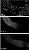
PGL-1 disappears from P granules during spermatogenesis. Immunofluorescence micrograph of a gonad arm dissected from an adult male. The gonad was fixed and stained with the DNA dye, DAPI, anti-PGL-1 antiserum and anti-α-tubulin. The stages of spermatogenesis are indicated in the top panel. PGL-1 disappears from P granules after the pachytene stage and before the formation of meiotic spindles. Scale bar: 10 μm.

References

    1. Altmann M, Sonenberg N, Trachsel H. Translation in Saccharomyces cerevisiae: initiation factor 4E-dependent cell-free system. Mol Cell Biol. 1989;19:4467–4472. - PMC - PubMed
    1. Barstead RJ, Waterston RH. The basal component of the nematode dense-body is vinculin. J Biol Chem. 1989;264:10177–10185. - PubMed
    1. Barton MK, Schedl TB, Kimble J. Gain-of-function mutations of fem-3, a sex-determination gene in Caenorhabditis elegans. Genetics. 1987;115:107–119. - PMC - PubMed
    1. Beanan MJ, Strome S. Characterization of a germ-line proliferation mutation in C. elegans. Development. 1992;116:755–766. - PubMed
    1. Brenner S. The genetics of Caenorhabditis elegans. Genetics. 1974;77:71–94. - PMC - PubMed

Publication types

MeSH terms