. 2001 Oct 15;15(20):2675-86.
doi: 10.1101/gad.924501.

FIH-1: a novel protein that interacts with HIF-1alpha and VHL to mediate repression of HIF-1 transcriptional activity

Affiliations

FIH-1: a novel protein that interacts with HIF-1alpha and VHL to mediate repression of HIF-1 transcriptional activity

P C Mahon et al. Genes Dev. .

Erratum in

Abstract

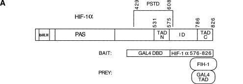
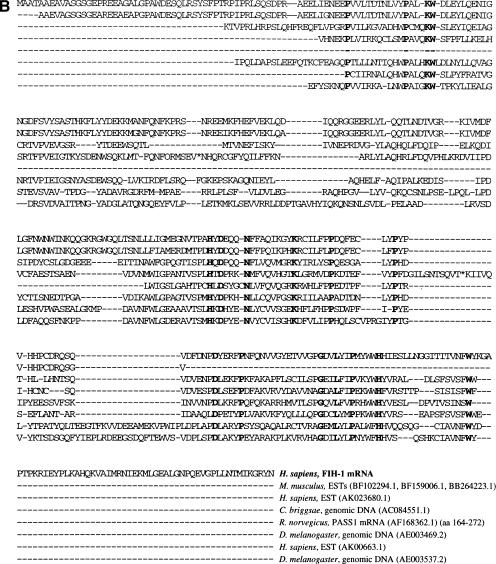
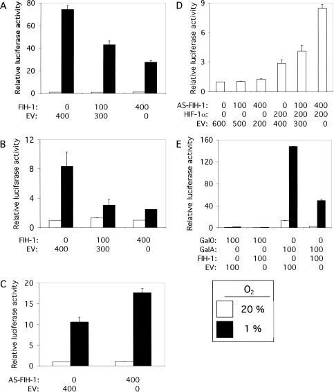
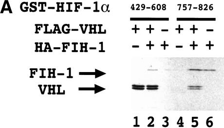
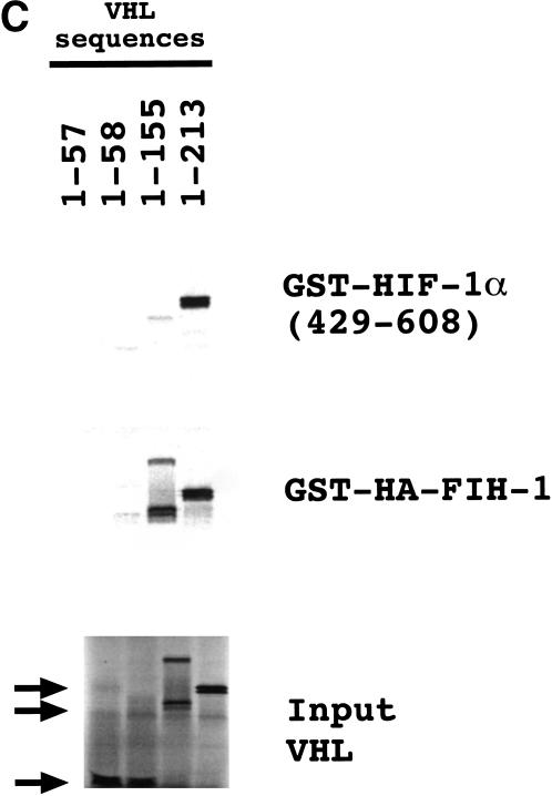
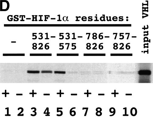
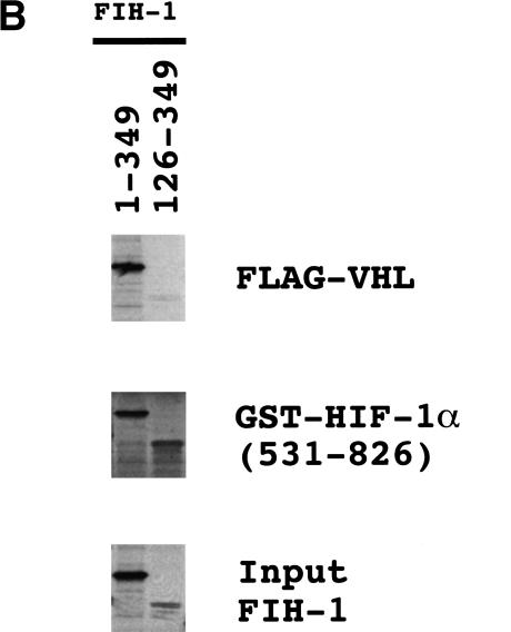
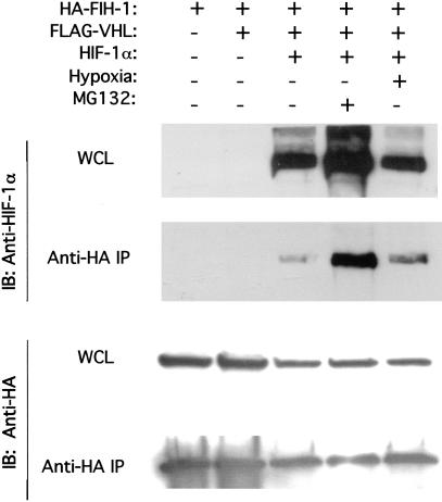
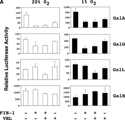
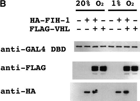
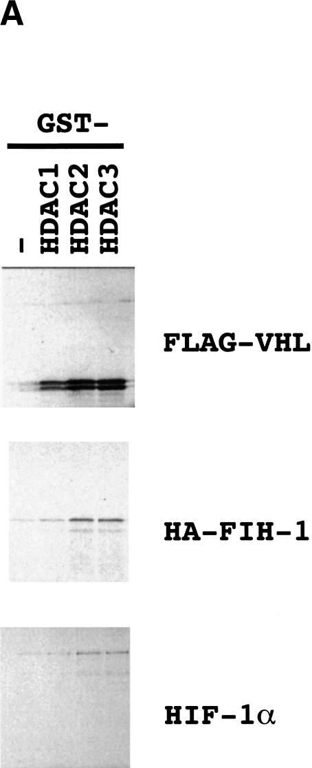
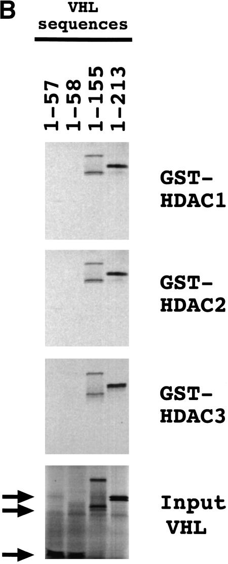
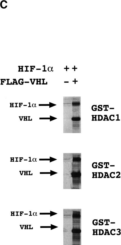
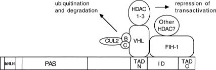
Hypoxia-inducible factor 1 (HIF-1) is a master regulator of oxygen homeostasis that controls angiogenesis, erythropoiesis, and glycolysis via transcriptional activation of target genes under hypoxic conditions. O(2)-dependent binding of the von Hippel-Lindau (VHL) tumor suppressor protein targets the HIF-1alpha subunit for ubiquitination and proteasomal degradation. The activity of the HIF-1alpha transactivation domains is also O(2) regulated by a previously undefined mechanism. Here, we report the identification of factor inhibiting HIF-1 (FIH-1), a protein that binds to HIF-1alpha and inhibits its transactivation function. In addition, we demonstrate that FIH-1 binds to VHL and that VHL also functions as a transcriptional corepressor that inhibits HIF-1alpha transactivation function by recruiting histone deacetylases. Involvement of VHL in association with FIH-1 provides a unifying mechanism for the modulation of HIF-1alpha protein stabilization and transcriptional activation in response to changes in cellular O(2) concentration.


Figures

Figure 1
Figure 1
Identification of FIH-1 by yeast two-hybrid screen. (A) Structure of HIF-1α and the bait and prey proteins. HIF-1α contains basic helix–loop–helix (bHLH) and PAS domains that are required for dimerization and DNA binding, a proline/serine/threonine-rich protein stabilization domain (PSTD), two transactivation domains (TAD-N and TAD-C), and an inhibitory domain (ID) that negatively regulates transactivation domain function under nonhypoxic conditions. For two-hybrid screening, the bait vector encoded a chimeric protein consisting of the DNA-binding domain from the yeast GAL4 transcription factor (GAL4 DBD) fused to residues 576–826 of HIF-1α. The prey vectors encoded the GAL4 transactivation domain (GAL4 TAD) fused to residues encoded by human brain cDNAs, one of which encoded FIH-1. (B) Amino acid sequence of FIH-1 and identification of related protein sequences. TBLASTN searches of GenBank databases were performed using the human FIH-1 sequence (top line). For each GenBank entry only those sequences showing significant similarity to FIH-1 are shown.
Figure 1
Figure 1
Identification of FIH-1 by yeast two-hybrid screen. (A) Structure of HIF-1α and the bait and prey proteins. HIF-1α contains basic helix–loop–helix (bHLH) and PAS domains that are required for dimerization and DNA binding, a proline/serine/threonine-rich protein stabilization domain (PSTD), two transactivation domains (TAD-N and TAD-C), and an inhibitory domain (ID) that negatively regulates transactivation domain function under nonhypoxic conditions. For two-hybrid screening, the bait vector encoded a chimeric protein consisting of the DNA-binding domain from the yeast GAL4 transcription factor (GAL4 DBD) fused to residues 576–826 of HIF-1α. The prey vectors encoded the GAL4 transactivation domain (GAL4 TAD) fused to residues encoded by human brain cDNAs, one of which encoded FIH-1. (B) Amino acid sequence of FIH-1 and identification of related protein sequences. TBLASTN searches of GenBank databases were performed using the human FIH-1 sequence (top line). For each GenBank entry only those sequences showing significant similarity to FIH-1 are shown.
Figure 2
Figure 2
Localization of HIF-1α residues interacting with FIH-1. (A) GST and GST–FIH-1 fusion proteins were expressed in E. coli, purified, incubated with 35S-labeled in vitro-translated HIF-1α, captured on glutathione–Sepharose beads, and analyzed by SDS-PAGE and autoradiography. (BD) GST-fusion proteins containing the indicated HIF-1α residues at their C terminus were incubated with 35S-labeled in vitro-translated FIH-1, captured on glutathione–Sepharose beads, and analyzed as described above. (0) GST only.
Figure 3
Figure 3
Effect of FIH-1 on HIF-1-mediated reporter gene transcription. Human 293 (A,D) or Hep3B (B,C) cells were cotransfected with pSV-Renilla, a reporter gene containing the SV40 promoter and Renilla luciferase-coding sequences, p2.1, a reporter gene containing a 68-bp hypoxia-response element upstream of the SV40-promoter and firefly luciferase-coding sequences, and the indicated amount of expression vector containing FIH-1 cDNA [inserted in either the sense or antisense (AS) orientation], HIF-1α cDNA, or empty vector (EV). For each expression vector, the amount (in nanograms) of plasmid DNA transfected is indicated. (E) Hep3B cells were co-transfected with pSV-Renilla, reporter pG5ElbLuc, containing five GAL4-binding sites upstream of an adenovirus Elb promoter and firefly luciferase coding sequences, expression vector encoding the GAL4 DNA-binding domain alone (Gal0) or fused to HIF-1α residues 531–826 (GalA), and EV or vector encoding FIH-1. In each panel, cells were exposed to 20% (open bars) or 1% (closed bars) O2 for 16 h and the ratio of firefly:Renilla luciferase activity was determined. The results were normalized to those for cells transfected with EV and exposed to 20% O2 (relative luciferase activity). The mean and standard deviation based on 3–9 independent transfections are shown.
Figure 4
Figure 4
Interaction of FIH-1, HIF-1α, and VHL in vitro. (A) GST-fusion proteins containing HIF-1α residues 429–608 or 757–826 were expressed in E. coli, purified, and incubated with 35S-labeled in vitro-translated FLAG-tagged VHL or HA-tagged FIH-1, captured on glutathione–Sepharose beads, and analyzed by SDS-PAGE and autoradiography. (B) 35S-labeled in vitro-translated FIH-1 residues 1–349 or 126–349 was incubated with unlabeled FLAG–VHL (top) or GST–HIF-1α(531–826) (middle), which were pulled down on beads containing anti-FLAG antibody or glutathione, respectively, and analyzed by SDS-PAGE along with aliquots of the input FIH-1 polypeptides (bottom). (C) 35S-labeled in vitro-translated FLAG–VHL truncated at its C terminus as indicated was incubated with unlabeled lysate-treated GST–HIF-1α(429–608) (top) or GST–HA–FIH-1 (middle), which were captured on glutathione–Sepharose beads and analyzed by SDS-PAGE along with aliquots of the input VHL polypeptides (bottom). (D) GST or the indicated GST–HIF-1α fusion protein was preincubated in reticulocyte lysate (odd-numbered lanes) or buffer (even-numbered lanes), incubated with 35S-labeled FLAG–VHL, captured on glutathione–Sepharose beads, and analyzed as described above.
Figure 4
Figure 4
Interaction of FIH-1, HIF-1α, and VHL in vitro. (A) GST-fusion proteins containing HIF-1α residues 429–608 or 757–826 were expressed in E. coli, purified, and incubated with 35S-labeled in vitro-translated FLAG-tagged VHL or HA-tagged FIH-1, captured on glutathione–Sepharose beads, and analyzed by SDS-PAGE and autoradiography. (B) 35S-labeled in vitro-translated FIH-1 residues 1–349 or 126–349 was incubated with unlabeled FLAG–VHL (top) or GST–HIF-1α(531–826) (middle), which were pulled down on beads containing anti-FLAG antibody or glutathione, respectively, and analyzed by SDS-PAGE along with aliquots of the input FIH-1 polypeptides (bottom). (C) 35S-labeled in vitro-translated FLAG–VHL truncated at its C terminus as indicated was incubated with unlabeled lysate-treated GST–HIF-1α(429–608) (top) or GST–HA–FIH-1 (middle), which were captured on glutathione–Sepharose beads and analyzed by SDS-PAGE along with aliquots of the input VHL polypeptides (bottom). (D) GST or the indicated GST–HIF-1α fusion protein was preincubated in reticulocyte lysate (odd-numbered lanes) or buffer (even-numbered lanes), incubated with 35S-labeled FLAG–VHL, captured on glutathione–Sepharose beads, and analyzed as described above.
Figure 5
Figure 5
Interaction of FIH-1 and HIF-1α in human cells. Human 293 cells were co-transfected with expression vectors encoding HA–FIH-1, FLAG–VHL, and HIF-1α, as indicated. The transfected cells were untreated, exposed to MG132, or subjected to hypoxia (1% O2) prior to lysis. Aliquots of whole cell lysate (WCL) and anti-HA immunoprecipitate (IP) were analyzed by immunoblot (IB) assay with antibodies that recognize HIF-1α (top) and HA (bottom).
Figure 6
Figure 6
Functional interaction of FIH-1 and VHL to repress HIF-1α transactivation domain function. (A) Hep3B cells were cotransfected with reporters pSV-Renilla and pG5E1bLuc, expression vector encoding the GAL4 DNA-binding domain alone (Gal0) or a GAL4–HIF-1α fusion protein, and expression vectors encoding no protein, FIH-1, or VHL. The GAL4-fusion proteins (containing the indicated HIF-1α residues) tested were GalA (531–826), GalG (757–826), GalL (531–575), and GalH (786–826). The relative luciferase activity represents the ratio of firefly:Renilla luciferase for each construct normalized to the result for Gal0. (B) Immunoblot analysis of lysates from transfected cells using monoclonal antibodies against the GAL4 DNA-binding domain (DBD), FLAG, and HA to detect expression of GalA (top), FLAG–VHL (middle), and HA–FIH-1 (bottom), respectively.
Figure 6
Figure 6
Functional interaction of FIH-1 and VHL to repress HIF-1α transactivation domain function. (A) Hep3B cells were cotransfected with reporters pSV-Renilla and pG5E1bLuc, expression vector encoding the GAL4 DNA-binding domain alone (Gal0) or a GAL4–HIF-1α fusion protein, and expression vectors encoding no protein, FIH-1, or VHL. The GAL4-fusion proteins (containing the indicated HIF-1α residues) tested were GalA (531–826), GalG (757–826), GalL (531–575), and GalH (786–826). The relative luciferase activity represents the ratio of firefly:Renilla luciferase for each construct normalized to the result for Gal0. (B) Immunoblot analysis of lysates from transfected cells using monoclonal antibodies against the GAL4 DNA-binding domain (DBD), FLAG, and HA to detect expression of GalA (top), FLAG–VHL (middle), and HA–FIH-1 (bottom), respectively.
Figure 7
Figure 7
Interaction of VHL and FIH-1 with histone deacetylases. (A) GST and GST–HDAC fusion proteins were incubated with 35S-labeled FLAG–VHL (top), HA–FIH-1 (middle), or HIF-1α (bottom), captured on glutathione–Sepharose beads, and analyzed by SDS-PAGE and autoradiography. (B) GST–HDAC fusion proteins were incubated with 35S-labeled FLAG–VHL truncated at its C terminus as indicated and analyzed as described above. (C) GST–HDAC fusion proteins were incubated with 35S-labeled FLAG–VHL and/or 35S-labeled HIF-1α.
Figure 7
Figure 7
Interaction of VHL and FIH-1 with histone deacetylases. (A) GST and GST–HDAC fusion proteins were incubated with 35S-labeled FLAG–VHL (top), HA–FIH-1 (middle), or HIF-1α (bottom), captured on glutathione–Sepharose beads, and analyzed by SDS-PAGE and autoradiography. (B) GST–HDAC fusion proteins were incubated with 35S-labeled FLAG–VHL truncated at its C terminus as indicated and analyzed as described above. (C) GST–HDAC fusion proteins were incubated with 35S-labeled FLAG–VHL and/or 35S-labeled HIF-1α.
Figure 8
Figure 8
Negative regulation of HIF-1α protein stability and transcriptional activity under nonhypoxic conditions mediated by VHL and FIH-1. Elongins B and C and cullin 2 are required for E3 ubiquitin–protein ligase activity, whereas HDACs repress transactivation.

References

    1. An WG, Kanekal M, Simon MC, Maltepe E, Blagosklonny MV, Neckers LM. Stabilization of wild-type p53 by hypoxia-inducible factor 1α. Nature. 1998;392:405–408. - PubMed
    1. Arany Z, Huang LE, Eckner R, Bhattacharya S, Jiang C, Goldberg MA, Bunn HF, Livingston DM. An essential role for p300/CBP in the cellular response to hypoxia. Proc Natl Acad Sci. 1996;93:12969–12973. - PMC - PubMed
    1. Bigner SH, Vogelstein B. Cytogenetics and molecular genetics of malignant gliomas and medulloblastoma. Brain Pathol. 1990;1:12–18. - PubMed
    1. Bruick RK. Expression of the gene encoding the proapoptotic Nip3 protein is induced by hypoxia. Proc Natl Acad Sci. 2000;97:9082–9087. - PMC - PubMed
    1. Carmeliet P, Dor Y, Herbert J-M, Fukumura D, Brusselmans K, Dewerchin M, Neeman M, Bono F, Abramovitch R, Maxwell P, et al. Role of HIF-1α in hypoxia-mediated apoptosis, cell proliferation, and tumour angiogenesis. Nature. 1998;394:485–490. - PubMed

Publication types

MeSH terms

Substances

Associated data