. 2001 Oct 15;15(20):2702-19.
doi: 10.1101/gad.915701.

Tissue-specific GATA factors are transcriptional effectors of the small GTPase RhoA

Affiliations

Tissue-specific GATA factors are transcriptional effectors of the small GTPase RhoA

F Charron et al. Genes Dev. .

Abstract

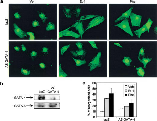
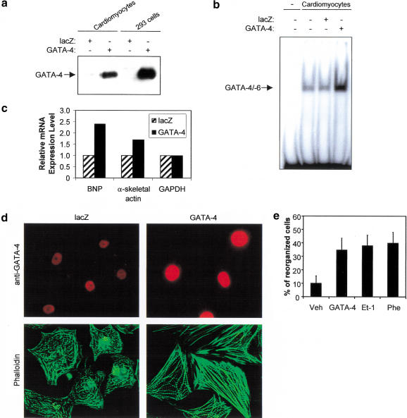
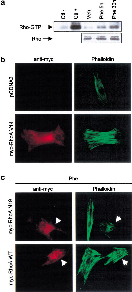
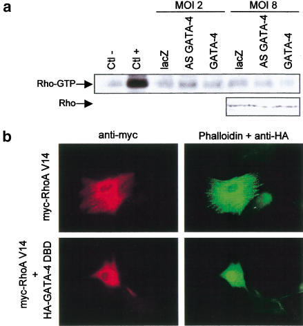
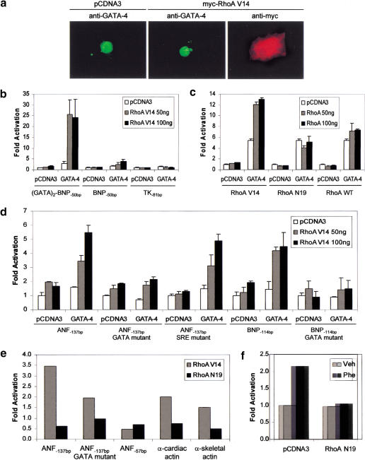
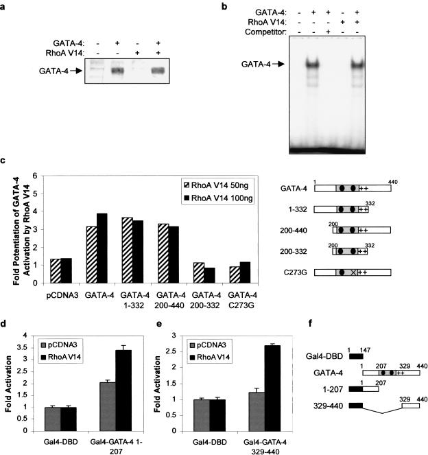
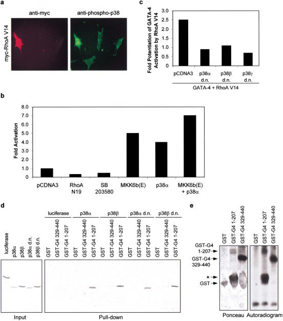
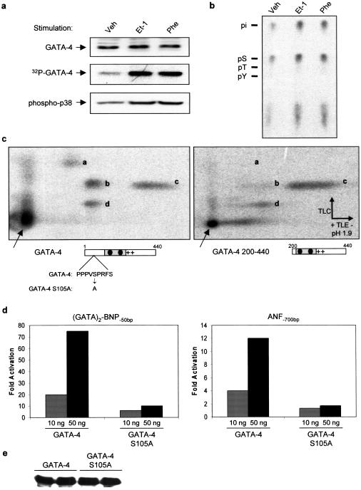
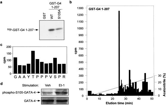
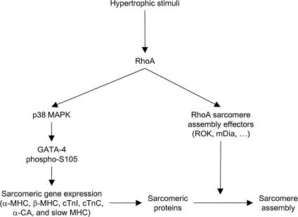
Rho-like GTPases play a pivotal role in the orchestration of changes in the actin cytoskeleton in response to receptor stimulation, and have been implicated in transcriptional activation, cell growth regulation, and oncogenic transformation. Recently, a role for RhoA in the regulation of cardiac contractility and hypertrophic cardiomyocyte growth has been suggested but the mechanisms underlying RhoA function in the heart remain undefined. We now report that transcription factor GATA-4, a key regulator of cardiac genes, is a nuclear mediator of RhoA signaling and is involved in the control of sarcomere assembly in cardiomyocytes. Both RhoA and GATA-4 are essential for sarcomeric reorganization in response to hypertrophic growth stimuli and overexpression of either protein is sufficient to induce sarcomeric reorganization. Consistent with convergence of RhoA and GATA signaling, RhoA potentiates the transcriptional activity of GATA-4 via a p38 MAPK-dependent pathway that phosphorylates GATA-4 activation domains and GATA binding sites mediate RhoA activation of target cardiac promoters. Moreover, a dominant-negative GATA-4 protein abolishes RhoA-induced sarcomere reorganization. The identification of transcription factor GATA-4 as a RhoA mediator in sarcomere reorganization and cardiac gene regulation provides a link between RhoA effects on transcription and cell remodeling.


Figures

Figure 1
Figure 1
GATA-4 is essential for Et-1 and Phe-induced cardiomyocyte sarcomere reorganization. (a) Cardiomyocytes infected with a lacZ control adenovirus (lacZ) or with the antisense GATA-4 adenovirus (AS GATA-4) were stimulated for 48 h with vehicle (Veh), Et-1, or Phe. Cardiomyocytes were fixed and actin filaments were revealed using phalloidin-FITC. (b) Western blot analysis of GATA-4 protein confirming that the GATA-4 antisense adenovirus specifically decreases GATA-4 protein levels. (c) Quantification of the percentage of reorganized cardiomyocytes. Cells were scored as described in Materials and Methods. The data are the average of two independent experiments.
Figure 2
Figure 2
Up-regulation of GATA-4 activity induces contractile protein gene expression and sarcomere reorganization in cardiomyocytes. (a) Western blot analysis of nuclear extracts from cardiomyocytes and 293 cells infected with a lacZ control or with GATA-4-expressing adenovirus. (b) Increased GATA DNA-binding activity in cardiomyocytes infected with the GATA-4 adenovirus. EMSAs were performed using nuclear extracts from cardiomyocytes infected with lacZ or GATA-4 adenovirus and the ANF −120-bp GATA probe. (c) Overexpression of GATA-4 induces cardiac gene expression. Total RNA (20 μg) extracted from cardiomyocytes infected with lacZ or GATA-4 adenovirus was analyzed by Northern blot and quantified by PhosphorImager, as described in Materials and Methods. (d) Up-regulation of GATA-4 activity induces cardiomyocyte sarcomere reorganization. Cardiomyocytes infected with lacZ or GATA-4 adenovirus were fixed and costained using phalloidin-FITC (green) and anti-GATA-4 antibody (red). (e) Quantification of the percentage of reorganized cardiomyocytes. Cells were scored as described in Material and Methods.
Figure 3
Figure 3
RhoA is required for Et-1- and Phe-induced cardiomyocyte sarcomere reorganization. (a) Phe induces Rho activity in a time-dependent manner. Whole-cell extracts were prepared from cardiomyocytes stimulated with Phe for 5 and 30 h. The active form of Rho (Rho-GTP) was selectively affinity-precipitated using a GST–Rhotekin protein and revealed using anti-Rho antibody. Extracts were incubated in presence of an excess of GDP (Ctl−) or GTP-γ-S (Ctl+) as negative and positive controls, respectively. The panel below shows total RhoA protein level as assessed by Western blot analysis. (b) RhoA V14 induces sarcomere reorganization in cardiomyocytes. Cardiomyocytes transfected with 2 μg of pCDNA3 or pCDNA3–myc–RhoA V14 were fixed and costained using phalloidin-FITC (green) and anti-myc antibody (red). (c) RhoA N19, but not RhoA WT, inhibits Phe-induced cardiomyocyte sarcomere reorganization. Cardiomyocytes transfected with 2 μg of pCDNA3–myc–RhoA WT or pCDNA3–myc–RhoA N19 were stimulated for 48 h with Phe, fixed, and costained using phalloidin-FITC (green) and anti-myc antibody (red). The arrow indicates myc-positive cardiomyocytes.
Figure 4
Figure 4
(a) Overexpression or down-regulation of GATA-4 does not affect RhoA activity in cardiomyocytes. Cardiomyocytes were infected with lacZ, AS GATA-4, or GATA-4 adenovirus, at MOIs of 2 or 8, for 48 h. Rho activity was assessed as described in Figure 3a. Extracts were incubated in presence of an excess of GDP (Ctl−) or GTP-γ-S (Ctl+) as negative and positive controls, respectively. The panel below is from a Western blot showing the level of total RhoA protein in the extracts. (b) GATA-4 activity is required for RhoA-induced sarcomeric reorganization. Cardiomyocytes were transiently transfected with myc–RhoA V14 in presence or absence of an expression vector encoding a HA-tagged dominant-negative GATA-4 protein lacking transcriptional activation domain (HA–GATA-4 DBD). Cells were fixed and costained with anti-myc antibody (red), anti-HA (nuclear green staining), and phalloidin-FITC (cytoplasmic green staining). Note how the presence of the GATA-4 DBD abolishes the effects of RhoA on cell size and sarcomeric reorganization.
Figure 5
Figure 5
RhoA potentiates GATA-4 transcriptional activity. (a) RhoA V14 does not affect GATA-4 protein level or subcellular localization. Cardiomyocytes transfected with 2 μg of pCDNA3 or pCDNA3–myc–RhoA V14 were fixed and costained using an anti-myc antibody (red) and an anti-GATA-4 antibody (green). (b) RhoA potentiates GATA-4 transcriptional activity. NIH 3T3 cells were transfected with 100 ng of pCDNA3 or pCDNA3–GATA-4 and increasing amounts of pCDNA3–myc–RhoA V14 (0, 50, and 100 ng), together with the indicated reporter plasmid. BNP−50bp and TK−81bp are the minimal BNP and thymidine kinase promoters cloned upstream of a luciferase reporter gene, respectively. (GATA)2–BNP−50bp contains two GATA elements from the BNP promoter in front of BNP−50bp. Reporter activity was assayed 48 h after transfection. (c) RhoA-mediated potentiation of GATA-4 transcriptional activation requires RhoA activity. NIH 3T3 cells were transfected with 100 ng of pCDNA3 or pCDNA3–GATA-4 and increasing amounts of pCDNA3–myc–RhoA V14, pCDNA3–myc–RhoA N19, or pCDNA3–myc–RhoA WT, together with the (GATA)2–BNP−50bp reporter plasmid. (d) RhoA potentiates GATA-4 activity on cardiac gene promoters. NIH 3T3 cells were transfected with 100 ng of pCDNA3 or pCDNA3–GATA-4 and increasing amounts of pCDNA3–myc–RhoA V14, together with the indicated reporter plasmid. The data shown in b–d are the mean ± S.D. of 4–6 independent determinations. (e) GATA elements mediate transcriptional regulation by RhoA in cardiomyocytes. ANF, α-cardiac actin, and α-skeletal actin–luciferase reporter constructs were cotransfected with 300 ng of RhoA expression vectors into primary neonate cardiomyocyte cultures. (f) Functional RhoA proteins are also required for transcriptional activation of the ANF promoter in response to Phe stimulation. The ANF-luciferase reporter (1.5 μg) was cotransfected with 1 μg of RhoA N19 or the corresponding empty vector (pCDNA3). Phe treatment was as described in Figure 1. The results in e and f are each from a representative experiment carried out in duplicate.
Figure 6
Figure 6
RhoA potentiates GATA-4 activity by stimulating the transcriptional activity of its activation domains. RhoA V14 does not affect (a) GATA-4 protein level or (b) GATA-4 DNA-binding activity, as assessed by Western blot and EMSA, using nuclear extracts of NIH 3T3 cells ectopically expressing either one or both proteins respectively. (c) The N- or C-terminal transactivation domain of GATA-4 is required for potentiation by RhoA. NIH 3T3 cells were transfected with 100 ng of pCDNA3 or pCDNA3–GATA-4 mutants and increasing amounts of pCDNA3–myc–RhoA V14, together with the (GATA)2–BNP−50bp reporter plasmid. The results are expressed as the ratio of the activity of the GATA-dependent reporter in presence of GATA-4 and RhoA proteins over the activity of the same reporter in presence of GATA-4 proteins alone. The GATA-4 mutants are depicted at right. (d) The N- and (e) C-terminal transactivation domains of GATA-4 are sufficient to support potentiation by RhoA. NIH 3T3 cells were transfected with 100 ng of pCMX–Gal4–DBD, pCMX–Gal4–GATA-4 1–207, or pCMX–Gal4–GATA-4 329–440 and 100 ng of pCDNA3 or pCDNA3–myc–RhoA V14, together with the (UAS)5–TK−81bp reporter plasmid. The Gal4–GATA-4 constructs are depicted in f. The data in c, d, and e are the mean ± S.D. of 2 to 3 experiments each carried out in duplicate.
Figure 7
Figure 7
RhoA potentiates GATA-4 transcriptional activity through a p38 MAPK-dependent mechanism. (a) RhoA V14 activates p38 MAPK in cardiomyocytes. Cardiomyocytes were transfected with 1 μg of pCDNA3–myc–RhoA V14. Twenty-four hours later, the cells were fixed and costained with anti-myc (red) and anti-phospho-p38 (green) antibodies. Note the induced nuclear and perinuclear phospho-p38 staining in RhoA V14 positive cells. (b) The MKK6/p38 MAPK pathway potentiates endogenous cardiac GATA-4 activity. Cardiomyocytes were transfected with 100 ng of pCDNA3 or activated MKK6 [MKK6b(e)] or p38α MAPK, together with the (GATA)2–BNP−50bp reporter plasmid. (c) Dominant-negative p38 constructs block the potentiation of GATA-4 transcriptional activity by RhoA. NIH 3T3 cells were transfected with 100 ng of pCDNA3 or pCDNA3–GATA-4, 100 ng of pCDNA3–myc–RhoA V14, various dominant-negative (d.n.) isoforms of p38 MAPK, together with the (GATA)2–BNP−50bp reporter plasmid. (d) p38 MAPKs interact with the N-terminal domain of GATA-4. Pull-down assays were performed by incubating GST, GST–GATA-4 1–207, or GST–GATA-4 329–440 with [35S]methionine-labeled luciferase or p38 MAPK isoforms. The complexes were washed and resolved by 10% SDS-PAGE. (e) p38α phosphorylates GATA-4 in vitro. Activated p38α adsorbed on agarose beads was incubated with GST, GST–GATA-4 1–207, or GST–GATA-4 329–440 recombinant proteins in the presence of [γ-32P]ATP. The reaction mixtures were resolved by SDS-PAGE and analyzed by autoradiography. The asterisk denotes a degradation product containing the GATA-4 protein.
Figure 8
Figure 8
The RhoA-activating agonists Et-1 and Phe induce GATA-4 phosphorylation in cardiomyocytes. (a) Cardiomyocytes were labeled with [32P]orthophosphate and stimulated for 1 h with Et-1 or Phe. GATA-4 was immunoprecipitated and the immune complex was resolved by SDS-PAGE (32P-GATA-4; middle). In parallel, nuclear extracts were prepared from non-radiolabeled cardiomyocytes and total GATA-4 protein levels were analyzed by Western blot (GATA-4; top). In addition, Western blot analysis of whole-cell extracts using a phospho-specific antibody showed that Et-1 and Phe induce p38 activation in cardiomyocytes (phospho-p38; bottom). (b) Et-1 and Phe induce GATA-4 phosphorylation on serine residues. Radiolabeled GATA-4 bands from a were excised and subjected to phosphoamino acid analysis, as described in Materials and Methods. Note that GATA-4 is phosphorylated almost exclusively on serine residues. (c) Phosphopeptide mapping analysis of 32P-labeled wild-type (left) and N-terminal deleted (right) GATA-4; note the absence of spot ‘a‘ when the N-terminal domain is removed. (d) A conserved MAPK phosphorylation site is essential for maximal GATA-4 transcriptional activity. Indicated amount of GATA-4 and GATA-4 S105A were transfected with the (GATA)2–BNP−50bp or the ANF−700bp reporter plasmids in NIH 3T3 cells and reporter activity was measured 48 h later. Note the drastic effect of the MAPK consensus site S105A mutation on GATA-4 transcriptional activity, even though both proteins are expressed at equal levels as shown by Western blot analysis (e).
Figure 9
Figure 9
p38 MAPK phosphorylates GATA-4 on Ser 105 in vitro. (a) Recombinant active p38α was incubated with GST, GST–GATA-4 1–207, or GST–GATA4 1–207 S105A in the presence of [γ-32P]ATP. The reaction products were analysed by SDS-PAGE and autoradiography. (b) The labeled GST–GATA-4 WT band from a was excised from the gel and digested with trypsin and endoproteinase GluC. The resulting peptides were separated by HPLC on a microbore C18 column developed with a nonlinear acetonitrile gradient. The fractions were collected manually and the radioactivity was measured by Cerenkov counting. (c) The major 32P-containing peptide in GST–GATA-4 1–207 was subjected to automatic Edman degradation. The amount of radioactivity released from each degradation cycle was determined by Cerenkov counting. The radioactivity in the first cycle corresponds to noncovalently bound 32P released by the TFA wash during the first cycle. (d) Western blot analysis of lysate proteins from cultured cardiomyocytes treated with vehicle (Veh) or ET-1 for 3 h using the phospho-S105 GATA-4 or the GATA-4 specific antibodies. Cardiomyocytes were infected with a GATA-4-expressing adenovirus to increase the signal intensity, as described in Liang et al. (2001). Note that Et-1 treatment increases the level of S105 phophorylated GATA-4.
Figure 10
Figure 10
Hypothetical model for the role of GATA-4 as an effector of RhoA and a regulator of cardiomyocyte sarcomere reorganization. Hypertrophic stimuli induce RhoA activity, which in turn potentiates GATA-4 transcriptional activity via p38 MAPK, leading to genetic reprogramming and induction of sarcomeric gene expression. p38 MAPK regulation of GATA-4 involves direct GATA-4 phosphorylation and possibly activation of GATA-4 cofactors like Mef2. RhoA, through activation of effectors such as mDia and ROK, induces polymerization of existing and newly synthesized sarcomeric proteins into contractile filaments, leading to sarcomere formation. Together, these two RhoA-dependent pathways would insure initiation and maintenance of the sarcomere reorganization and the hypertrophic state.

References

    1. Acuto O, Cantrell D. T cell activation and the cytoskeleton. Annu Rev Immunol. 2000;18:165–184. - PubMed
    1. Andreka P, Zang J, Dougherty C, Slepak TI, Webster KA, Bishopric NH. Cytoprotection by Jun kinase during nitric oxide-induced cardiac myocyte apoptosis. Circ Res. 2001;88:305–312. - PubMed
    1. Aoki H, Izumo S, Sadoshima J. Angiotensin II activates RhoA in cardiac myocytes: A critical role of RhoA in angiotensin II-induced premyofibril formation. Circ Res. 1998;82:666–676. - PubMed
    1. Argentin S, Ardati A, Tremblay S, Lihrmann I, Robitaille L, Drouin J, Nemer M. Developmental stage-specific regulation of atrial natriuretic factor gene transcription in cardiac cells. Mol Cell Biol. 1994;14:777–790. - PMC - PubMed
    1. Bar-Sagi D, Hall A. Ras and Rho GTPases: A family reunion. Cell. 2000;103:227–238. - PubMed

Publication types

MeSH terms