

Proton chemical shift imaging in normal pressure hydrocephalus
- PMID: 11673158
- PMCID: PMC7974445
Proton chemical shift imaging in normal pressure hydrocephalus
Abstract
Background and purpose: Differentiation of normal pressure hydrocephalus (NPH) from other types of dementia and the selection of appropriate candidates for shunt surgery remain a clinical challenge. The aims of this study were to assess the efficacy of cerebral metabolites depicted by proton chemical shift imaging (1H-CSI) in distinguishing NPH from other dementias and to examine the relationship between metabolite changes and the outcome of shunt surgery.
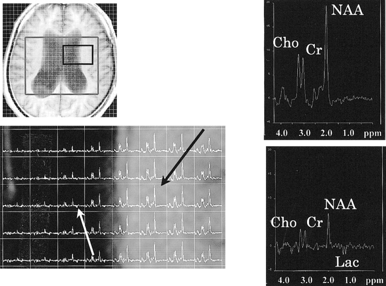
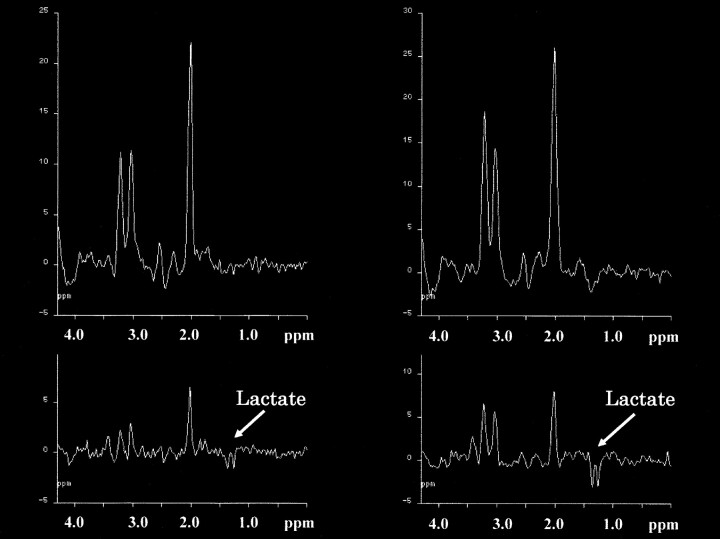
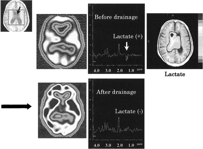
Methods: 1H-CSI measurements were obtained in nine patients with clinical diagnosis of NPH; six patients with other types of dementia, including Alzheimer and Pick disease; and five control subjects. The 1H-CSI sequence consisted of a double spin-echo sequence with imaging parameters of 2000/135/4-2 (TR/TE/acquisitions). Volumes of interest were selected from a section through the lateral ventricles. The peak areas and ratios of N-acetylaspartate, creatine, choline, and lactate were calculated. In two patients, follow-up 1H-CSI and N-isopropyl (123I)-p-iodoamphetamine brain perfusion imaging were available after treatment with continuous spinal drainage.
Results: Lactate peaks were observed in the lateral ventricles for all patients with NPH (lactate/creatine, 0.23 +/- 0.14) but not for patients with other types of dementia or control subjects. In all cases, we noted no significant differences in the peak ratios in the voxels located at the white matter near the lateral ventricles. In one patient with NPH, intraventricular lactate disappeared and regional CBF recovered after drainage.
Conclusion: The intraventricular lactate level may be useful in differentiating NPH from other types of dementia.
Figures
Comment in
-
Normal pressure hydrocephalus and deep white matter ischemia: which is the chicken, and which is the egg?AJNR Am J Neuroradiol. 2001 Oct;22(9):1638-40. AJNR Am J Neuroradiol. 2001. PMID: 11673152 Free PMC article. No abstract available.
-
Ventricular lactate in normal pressure hydrocephalus: from where has it come to where does it go?AJNR Am J Neuroradiol. 2002 Jun-Jul;23(6):1061; author reply 1061-2. AJNR Am J Neuroradiol. 2002. PMID: 12063242 Free PMC article. No abstract available.
References
-
- Adams RD, Fisher CM, Hakim S, et al. Symptomatic occult hydrocephalus with “normal” cerebrospinal fluid pressure. N Engl J Med 1965;273:117-126 - PubMed
-
- Ojemann RG. Normal pressure hydrocephalus. Clin Neurosurg 1970;18:337-370 - PubMed
-
- Masters JC, O'Grady M. Normal pressure hydrocephalus. A potentially reversible form of dementia. J Psychosoc Nurs Ment Health Serv 1992;30:25-28 - PubMed
-
- Benson DF, LeMay M, Patten DH, Rubens AB. Diagnosis of normal-pressure hydrocephalus. N Engl J Med 1970;283:609-615 - PubMed
MeSH terms
Substances
LinkOut - more resources
Full Text Sources
Medical