

Restricted diffusion within ring enhancement is not pathognomonic for brain abscess
- PMID: 11673170
- PMCID: PMC7974430
Restricted diffusion within ring enhancement is not pathognomonic for brain abscess
Abstract
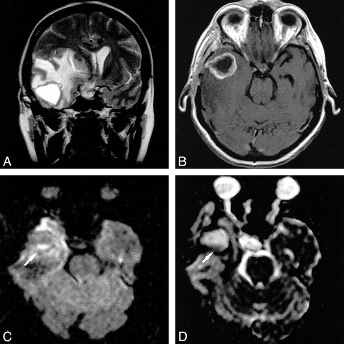
Background and purpose: Recent experience suggests that diffusion-weighted MR imaging may be decisive in the differential diagnosis of ring-enhancing cerebral lesions. Whether restricted diffusion within a ring-enhancing cerebral mass lesion is pathognomonic for abscess was studied.
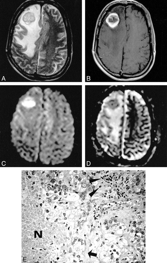
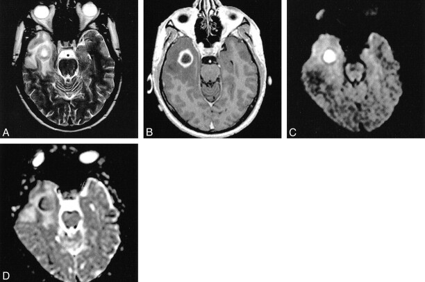
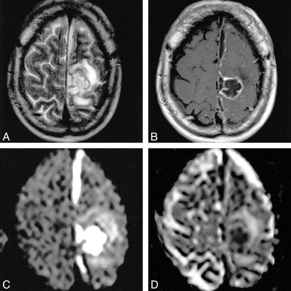
Methods: Seventeen patients with ring-enhancing cerebral lesions (three abscesses, six glioblastomas, eight metastases) on conventional contrast-enhanced T1-weighted images were examined with echo-planar diffusion-weighted MR imaging. Apparent diffusion coefficient (ADC) maps and the ADCs were calculated for all lesions. Lesions with signs of intralesional hemorrhage on unenhanced T1-weighted images were excluded.
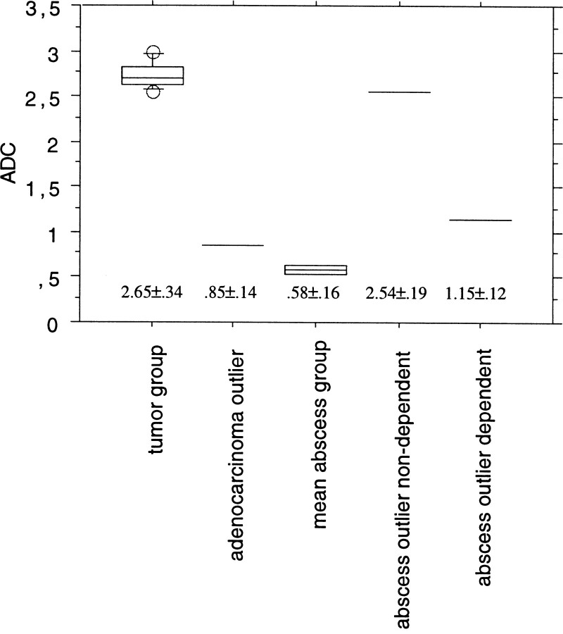
Results: The central portion of all six glioblastomas and seven of eight metastases showed unrestricted diffusion, whereas two of three abscesses showed restricted diffusion (low ADC values) in their cavity. However, restricted diffusion also was found in one metastasis, and one abscess within a postoperative cavity showed unrestricted diffusion within a larger nondependent portion.
Conclusion: In patients with ring-enhancing cerebral mass lesions, restricted diffusion might be characteristic but is not pathognomonic for abscess, as low ADC values also may be found in brain metastases.
Figures
References
-
- Ebisu T, Tanaka C, Umeda M, et al. Discrimination of brain abscess from necrotic or cystic tumors by diffusion-weighted echo planar imaging. Magn Reson Imaging 1996;14:1113-1116 - PubMed
-
- Kim YJ, Chang K-H, Song IC, et al. Brain abscess and necrotic or cystic brain tumor: discrimination with signal intensity on diffusion-weighted MR imaging. AJR Am J Roentgenol 1998;171:1487-1490 - PubMed
-
- Noguchi K, Watanabe N, Nagayoshi T, et al. Role of diffusion-weighted echo-planar MRI in distinguishing between brain abscess and tumour: a preliminary report. Neuroradiology 1999;41:171-174 - PubMed
-
- Tsuchiya K, Yamakami N, Hachiya J, Saito I, Kobayashi H. Multiple brain abscesses: differentiation from cerebral metastases by diffusion-weighted magnetic resonance imaging. Int J Neuroradiol 1998;4:258-262
MeSH terms
LinkOut - more resources
Full Text Sources
Medical