

Expression of chemokine receptors in vernal keratoconjunctivitis
- PMID: 11673306
- PMCID: PMC1723775
- DOI: 10.1136/bjo.85.11.1357
Expression of chemokine receptors in vernal keratoconjunctivitis
Abstract
Background/aims: Chemokines are small peptides which are potent activators and chemoattractants for leucocyte subpopulations. Their action is mediated by a family of seven transmembrane spanning G-protein coupled receptors. The aims of this study were to examine the expression of the chemokine receptors CCR1, CCR3, CCR5, CXCR3, and CXCR4 in the conjunctiva of patients with vernal keratoconjunctivitis (VKC) and to investigate the phenotype of inflammatory cells expressing these chemokine receptors.
Methods: Conjunctival biopsy specimens from 16 patients with active VKC, and eight control subjects were studied by immunohistochemical techniques using a panel of monoclonal antibodies directed against human CCR1, CCR3, CCR5, CXCR3, and CXCR4. The phenotype of inflammatory cells expressing chemokine receptors was examined by double immunohistochemistry.
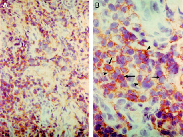
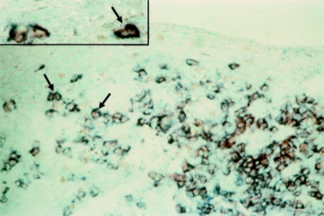
Results: In the normal conjunctiva, few inflammatory cells expressed CXCR3 in five of eight specimens. There was no immunoreactivity for CCR1, CCR3, CCR5, and CXCR4. In VKC specimens, membranous immunoreactivity for CXCR3 was noted on inflammatory cells in all specimens. Compared with control specimens, VKC specimens showed significantly more inflammatory cells expressing CXCR3 (54.3 (SD 34.3) v 3.3 (5.0); p<0.001). Few CCR1+, CCR3+, CCR5+, and CXCR4+ inflammatory cells were observed in only three of 16 specimens. Double immunohistochemistry revealed that all CXCR3 positive inflammatory cells were CD3 positive T lymphocytes and that 61.7% (3.7%) of the infiltrating T lymphocytes were reactive for CXCR3.
Conclusions: CXCR3 is the predominant chemokine receptor and is expressed abundantly on T lymphocytes in the conjunctiva of patients with active VKC. These data suggest a potential role for CXCR3 receptors in the regulation of lymphocyte recruitment within conjunctiva of VKC patients. New therapeutic strategies that block CXCR3 may inhibit T lymphocyte recruitment and suppress adverse inflammatory reactions.
Figures
Similar articles
-
Expression of T lymphocyte chemoattractants and activation markers in vernal keratoconjunctivitis.Br J Ophthalmol. 2002 Oct;86(10):1175-80. doi: 10.1136/bjo.86.10.1175. Br J Ophthalmol. 2002. PMID: 12234902 Free PMC article.
-
Langerhans' cells in vernal keratoconjunctivitis express the costimulatory molecule B7-2 (CD86), but not B7-1 (CD80).Eye (Lond). 2001 Oct;15(Pt 5):648-54. doi: 10.1038/eye.2001.202. Eye (Lond). 2001. PMID: 11702979
-
The T-lymphocyte chemoattractant Mig is highly expressed in vernal keratoconjunctivitis.Am J Ophthalmol. 2003 Nov;136(5):853-60. doi: 10.1016/s0002-9394(03)00446-x. Am J Ophthalmol. 2003. PMID: 14597036
-
Immunopathogenesis of vernal keratoconjunctivitis.Bull Soc Belge Ophtalmol. 1996;261:15-24. Bull Soc Belge Ophtalmol. 1996. PMID: 9009358 Review.
-
Targeting of Th1-associated chemokine receptors CXCR3 and CCR5 as therapeutic strategy for inflammatory diseases.Mini Rev Med Chem. 2007 Nov;7(11):1089-96. doi: 10.2174/138955707782331768. Mini Rev Med Chem. 2007. PMID: 18045212 Review.
Cited by
-
T-cell characterization in chronic allergic eye disease.Curr Allergy Asthma Rep. 2003 Jul;3(4):358-62. doi: 10.1007/s11882-003-0099-1. Curr Allergy Asthma Rep. 2003. PMID: 12791216 Review.
-
Basic science and pathophysiology of ocular allergy.Curr Allergy Asthma Rep. 2004 Jul;4(4):326-31. doi: 10.1007/s11882-004-0079-0. Curr Allergy Asthma Rep. 2004. PMID: 15175149 Review.
-
An imbalance between frequency of CD4+CD25+FOXP3+ regulatory T cells and CCR4+ and CCR9+ circulating helper T cells is associated with active perennial allergic conjunctivitis.Clin Dev Immunol. 2013;2013:919742. doi: 10.1155/2013/919742. Epub 2013 Dec 4. Clin Dev Immunol. 2013. PMID: 24368924 Free PMC article.
-
Early inflammatory markers in elicitation of allergic contact dermatitis.BMC Dermatol. 2002 Aug 7;2:9. doi: 10.1186/1471-5945-2-9. BMC Dermatol. 2002. PMID: 12167174 Free PMC article. Clinical Trial.
-
Altered regulation of CXCR4 expression during aging contributes to increased CXCL12-dependent chemotactic migration of CD4(+) T cells.Aging Cell. 2012 Aug;11(4):651-8. doi: 10.1111/j.1474-9726.2012.00830.x. Epub 2012 Jun 4. Aging Cell. 2012. PMID: 22568557 Free PMC article.
References
Publication types
MeSH terms
Substances
LinkOut - more resources
Full Text Sources