

Entrance dose measurements for in-vivo diode dosimetry: Comparison of correction factors for two types of commercial silicon diode detectors
- PMID: 11674824
- PMCID: PMC5726170
- DOI: 10.1120/jacmp.v1i3.2642
Entrance dose measurements for in-vivo diode dosimetry: Comparison of correction factors for two types of commercial silicon diode detectors
Abstract
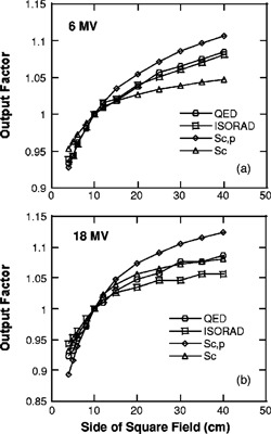
Silicon diode dosimeters have been used routinely for in-vivo dosimetry. Despite their popularity, an appropriate implementation of an in-vivo dosimetry program using diode detectors remains a challenge for clinical physicists. One common approach is to relate the diode readout to the entrance dose, that is, dose to the reference depth of maximum dose such as d(max) for the 10x10 cm(2) field. Various correction factors are needed in order to properly infer the entrance dose from the diode readout, depending on field sizes, target-to-surface distances (TSD), and accessories (such as wedges and compensate filters). In some clinical practices, however, no correction factor is used. In this case, a diode-dosimeter-based in-vivo dosimetry program may not serve the purpose effectively; that is, to provide an overall check of the dosimetry procedure. In this paper, we provide a formula to relate the diode readout to the entrance dose. Correction factors for TSD, field size, and wedges used in this formula are also clearly defined. Two types of commercial diode detectors, ISORAD (n-type) and the newly available QED (p-type) (Sun Nuclear Corporation), are studied. We compared correction factors for TSDs, field sizes, and wedges. Our results are consistent with the theory of radiation damage of silicon diodes. Radiation damage has been shown to be more serious for n-type than for p-type detectors. In general, both types of diode dosimeters require correction factors depending on beam energy, TSD, field size, and wedge. The magnitudes of corrections for QED (p-type) diodes are smaller than ISORAD detectors.
Figures
References
-
- Fletcher G. H., Textbook of Radiation Therapy, 3rd ed. (Lea and Febiger, Philadalphia, 1981).
-
- Essers M. and Mijnheer B. J., “In vivo dosimetry during external beam photon beam radiotherapy,” Int. J. Radiat. Oncol., Biol., Phys. 43, 245–259 (1999). - PubMed
-
- Alecu R., Alecu M., and Ochran T. G., “A method to improve the effectiveness of diode in vivo dosimetry,” Med. Phys. 25, 746–749 (1998). - PubMed
-
- Fontenla D. P., Curran J., Yaparpalvi R., and Vikram B., “Customization of a radiation management system to support in vivo patient dosimetry using diodes,” Med. Phys. 23, 1425–1429 (1996). - PubMed
-
- Fontenla D. P., Yaparpalvi R., Chui C.‐S., and Briot E., “The use of diode dosimetry in quality improvement of patient care in radiation therapy,” Med. Dosim 21, 235–241 (1996). - PubMed
Publication types
MeSH terms
Substances
LinkOut - more resources
Full Text Sources
