. 2002 Jan 4;277(1):763-73.
doi: 10.1074/jbc.M109134200. Epub 2001 Oct 23.

Convergence of multiple autophagy and cytoplasm to vacuole targeting components to a perivacuolar membrane compartment prior to de novo vesicle formation

Affiliations

Convergence of multiple autophagy and cytoplasm to vacuole targeting components to a perivacuolar membrane compartment prior to de novo vesicle formation

John Kim et al. J Biol Chem. .

Abstract

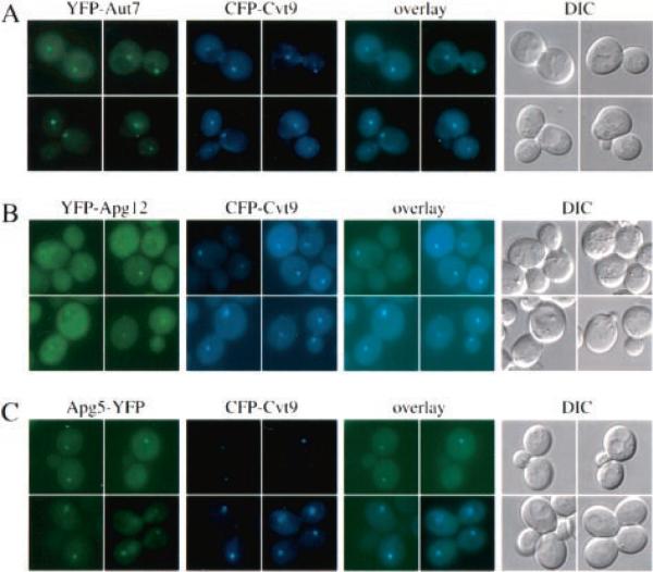
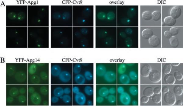
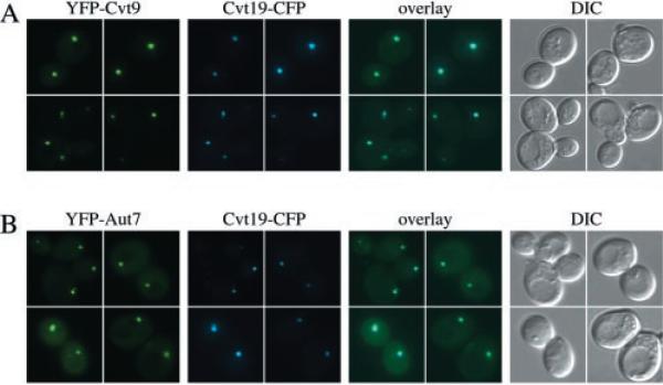
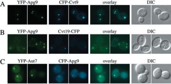
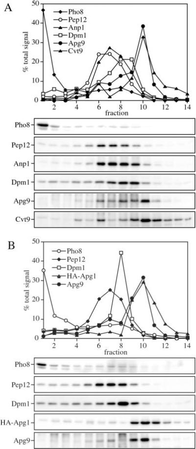
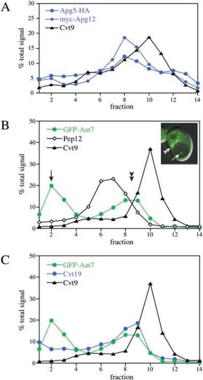
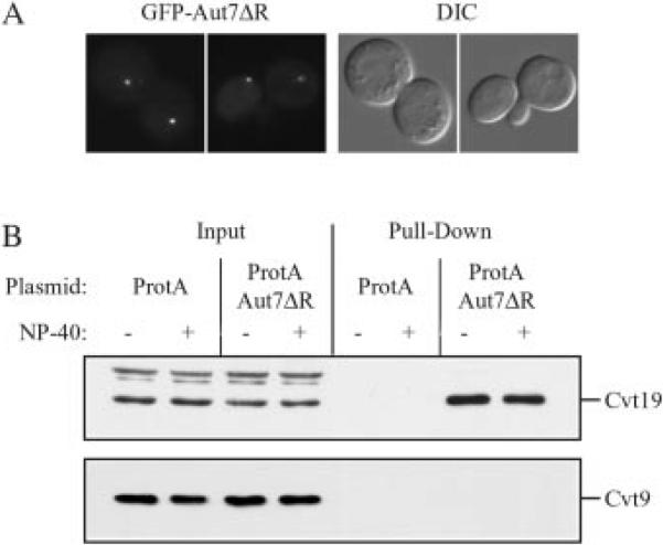
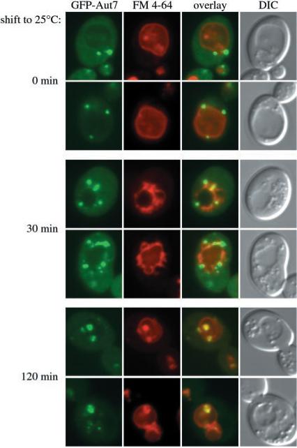
Under starvation conditions, the majority of intracellular degradation occurs at the lysosome or vacuole by the autophagy pathway. The cytoplasmic substrates destined for degradation are packaged inside unique double-membrane transport vesicles called autophagosomes and are targeted to the lysosome/vacuole for subsequent breakdown and recycling. Genetic analyses of yeast autophagy mutants, apg and aut, have begun to identify the molecular machinery as well as indicate a substantial overlap with the biosynthetic cytoplasm to vacuole targeting (Cvt) pathway. Transport vesicle formation is a key regulatory step of both pathways. In this study, we characterize the putative compartment from which both autophagosomes and the analogous Cvt vesicles may originate. Microscopy analyses identified a perivacuolar membrane as the resident compartment for both the Apg1-Cvt9 signaling complex, which mediates the switching between autophagic and Cvt transport, and the autophagy/Cvt-specific phosphatidylinositol 3-kinase complex. Furthermore, the perivacuolar compartment designates the initial site of membrane binding by the Apg/Cvt vesicle component Aut7, the Cvt cargo receptor Cvt19, and the Apg conjugation machinery, which functions in the de novo formation of vesicles. Biochemical isolation of the vesicle component Aut7 and density gradient analyses recapitulate the microscopy findings although also supporting the paradigm that components required for vesicle formation and packaging concentrate at subdomains within the donor membrane compartment.


Figures

Fig. 1
Fig. 1. Cvt9, the vesicle component Aut7, and the Apg12-Apg5 conjugation components co-localize by fluorescent microscopy
Wild type (SEY6210) (A and B) or aut7Δ (WPHYD7) (C) cells were co-transformed with the following pairs of copper-inducible plasmids. A, YFP-Aut7 (pCuYFPAUT7(426)) and CFP-Cvt9 (pCuCFPCVT9(424)); B, YFP-Apg12 (pCuYFPAPG12(426)) and CFP-Cvt9; C, Apg5-YFP (pAPG5YFP(426)) and CFP-Cvt9. The transformed cells were grown to midlog stage in SMD, induced with 30 μm CuSO4 for 2 h (A and B) or with 5 μm CuSO4 for 30 min (C), and examined with a Nikon E-800 fluorescence microscope equipped with a Hamamatsu Orca 2 digital camera and processed with Openlab software. The YFP fusions to the vesicle component Aut7 and the conjugation components Apg12 and Apg5 co-localize with CFP-Cvt9. The DIC panels are images obtained with differential interference contrast optics.
Fig. 2
Fig. 2. The Apg1 protein kinase and the Apg14 component of the PI 3-kinase co-localize with Cvt9
Wild type cells (SEY6210) were co-transformed with plasmids, under CUP1 copper-inducible control, expressing either YFP-Apg1 (pCuYFPAPG1(426)) and CFP-Cvt9 (pCuCFPCVT9(424)) (A) or YFP-Apg14 (pCuYFPAPG14(426)) and CFP-Cvt9 (B). Transformed cells were grown to midlog stage in SMD, induced for 1–2 h with 30 μm CuSO4, and viewed directly (B) or shifted to SD-N medium for 1 h prior to microscopy analysis (A). Images were captured and analyzed as in Fig. 1. The YFP fusion of the Apg1 kinase, and Apg14, a subunit of the Apg/Cvt-specific Vps15/34 PI-3 kinase complex, co-localize with the perivacuolar CFP-Cvt9 fusion protein. DIC, differential interference contrast.
Fig. 3
Fig. 3. Cvt9 co-localizes with the Cvt19 cargo receptor by fluorescence microscopy
Wild type cells (SEY6210) were co-transformed with the following pairs of plasmids. A, copper-inducible YFP-Cvt9 (pCuYFP-CVT9(426)) and Cvt19-CFP under endogenous promoter control (pCVT19CFP(414)); B, copper-inducible YFP-Aut7 (pCuYFPAUT7(426)) and Cvt19-CFP under endogenous promoter control (pCVT19CFP(414)). The transformed cells were grown to midlog stage in SMD, induced with 10 μm CuSO4 for 2 h, and examined with a Nikon E-800 fluorescence microscope as described in Fig. 1 and under “Experimental Procedures.” A fluorescent protein fusion of Cvt19 co-localizes with both Cvt9 and Aut7 at the perivacuolar compartment. DIC, differential interference contrast.
Fig. 4
Fig. 4. Cvt9 and Apg9 co-localize by fluorescent microscopy
Wild type cells (SEY6210) were co-transformed with plasmids, under CUP1 copper-inducible control, expressing either YFP-Apg9 (pCuYFPAPG9(426)) and CFP-Cvt9 (pCuCFPCVT9(424)) (A), YFP-Apg9 and Cvt19-CFP (pCVT19CFP(414)) (B), or YFP-Aut7 (pCuYFPAUT7(426)) and CFP-Apg9 (pCuCFPAPG9(424)) (C). Transformed cells were grown to midlog stage in SMD and induced for 1–2 h with 30 μm CuSO4. Images were taken and examined with a Nikon E-800 fluorescence microscope as described in Fig. 1 and under “Experimental Procedures.” Apg9 displays multiple punctate dots when co-expressed with Cvt19 and Aut7 but only a single dot when co-expressed with Cvt9. The YFP-Apg9 and CFP-Cvt9 dots co-localize. DIC, differential interference contrast.
Fig. 5
Fig. 5. Cvt9, Apg9, and Apg1 co-localize with each other but not with other endomembrane markers by density gradient separation
Subcellular co-localization of Apg9 and Cvt9 (A) and HA-Apg1 and Apg9 (B) by OptiPrep density gradients. The wild type strain (SEY6210) was co-transformed with the multicopy APG9 plasmid (pAPG9(426)) and the copper-inducible CVT9 plasmid (pCuCVT9(416)) (A) or a plasmid expressing an HA epitope-tagged Apg1 (pHAAPG1(423)) the multicopy APG9 plasmid (pAPG9(426)) (B). Cells were grown to midlog stage in SMD, and those in A incubated with 50 μm CuSO4 for 2 h to induce Cvt9 expression. The cells were converted to spheroplasts and lysed in PS200 buffer as described under “Experimental Procedures.” A total membrane fraction was isolated by centrifugation at 100,000 × g for 20 min and loaded to the top of a 10-ml OptiPrep linear gradient (10–55%). Following centrifugation at 100,000 × g for 12 h at 4 °C, 14 fractions were collected and analyzed by immunoblots with antiserum or antibodies to Pho8 (vacuole), Pep12 (endosome), Anp1 (cis-Golgi), Dpm1 (ER), Cvt9, Apg9, and the HA epitope (Apg1) as indicated. Indirect chemiluminescent detection and quantification of relative protein concentrations were performed with the Bio-Rad Fluor-S Max Imager. Cvt9, Apg1, and Apg9 co-localize to a dense part of the gradient and are separated from known endomembrane markers.
Fig. 6
Fig. 6. Co-localization analyses of Cvt9, the Apg conjugation proteins, and the vesicle components Aut7 and Cvt19
A, linear OptiPrep density gradient profiles of Apg conjugation components. Wild type cells (SEY6210) were co-transformed with plasmids expressing an HA-epitope fusion to Apg5 (pAPG5HA(424)), Myc-epitope fusion to Apg12 (pMyc-APG12(426)), and copper-inducible Cvt9 (pCuCVT9(415)). Transformed cells were grown to midlog in SMD and induced with 50 mm CuSO4 for 2 h to induce Cvt9 expression. A total membrane fraction was resolved by linear OptiPrep gradients (10–55%), and the collected fractions were analyzed by quantitative immunoblots with antiserum to Cvt9 and the HA (Apg5) and Myc (Apg12) epitopes as described in Fig. 5 and under “Experimental Procedures.” The Apg5 and Apg12 conjugation components can be resolved from Cvt9 by density gradient separation. B and C, linear density gradient profiles of GFP-Aut7, Cvt9, and Cvt19. The pep4Δ strain (TVY1) was co-transformed with copper-inducible plasmids expressing GFP-Aut7 (pCuGFPAUT7(416) and Cvt9 (pCuCVT9(414)). The transformed strain was grown to midlog stage in SMD and incubated with 20 μm CuSO4 for 2 h to induce GFP-Aut7 and Cvt9 expression. Just prior to subjecting the cells to density gradient analysis, the distribution of GFP-Aut7 was examined with a fluorescence microscope (see inset) as described under “Experimental Procedures.” The induced cells were then converted to spheroplasts, osmotically lysed, and a total membrane fraction was obtained and resolved on a 10–55% OptiPrep density gradient as described above. The collected gradient fractions were analyzed and quantified after immunoblotting with antibodies or antiserum to Aut7, Pep12, and Cvt9 (B), and Aut7, Cvt19, and Cvt9 (C). The quantification graphs in B and C are from the same gradient but are presented separately for clarity. Aut7 displays a bimodal distribution and co-fractionates with Cvt19 in the higher density peak. Both Aut7 and Cvt19 peak in a fraction that is resolved from Cvt9.
Fig. 7
Fig. 7. Cvt19 is co-isolated with protein A-Aut7ΔR
A, plasmid GFP-AUT7ΔR encoding GFP fused to Aut7 lacking the C-terminal arginine residue was transformed into the apg1Δ aut2Δ double knockout strain (WPHYD102). Cells were grown to midlog phase and examined by fluorescence microscopy as described under “Experimental Procedures.” GFP-Aut7ΔR localizes to a perivacuolar punctate dot. B, the protein A control vector (ProtA) or protein A-Aut7ΔR encoding plasmid (ProtA-AUT7ΔR) was co-transformed into the apg1Δ aut2Δ double knockout strain (WPHYD102) with one of the following plasmids: pCuCVT9(424), pAPG9(424), or pHA-APG12(424). Cells were collected either directly from midlog phase culture or after a 2.5-h induction with 20 μm CuSO4 to induce Cvt9 expression and converted to spheroplasts. Spheroplasts were then lysed in PBS lysis buffer in the absence or presence of 0.5% Nonidet P-40 (NP-40) as indicated. Lysates were incubated with Dynabeads M-500 cross-linked to human IgG. Protein A-Aut7ΔR and associated proteins were isolated by collecting Dynabeads from the crude cell lysates by magnetic isolation (Pull-Down). Proteins were resolved by SDS-PAGE and detected by immunoblot with antiserum against Cvt19, Cvt9, Apg9, or the HA epitope (Apg12). The positions of Cvt19 and Cvt9 are indicated. Input lanes corresponded to 2.5% of cell lysates used for each pull-down reaction. Protein A-Aut7ΔR pulls down Cvt19 but not Cvt9. The protein A-Aut7ΔR construct also pulled down HA-Apg12 but not Apg9 (data not shown, see text). DIC, differential interference contrast.
Fig. 8
Fig. 8. The perivacuolar compartment is a physiological intermediate in the Cvt and autophagy pathways
The apg9Δ pep4Δ strain was co-transformed with a plasmid expressing a temperature conditional allele of Apg9 (pAPG9ts(414)) and the copper-inducible GFP-Aut7 fusion protein (pCuGFPAUT7(416)). The transformed cells were grown at nonpermissive temperature (37 °C) to midlog stage, induced with 10 μm CuSO4 for 2 h, and labeled with FM 4–64 at 37 °C. The cultures were then incubated in SD-N medium for 1 h at 37 °C and viewed directly or shifted to permissive temperature (25 °C) for 30 and 120 min prior to fluorescence microscopy analysis as described in Fig. 1 and under “Experimental Procedures.” GFP-Aut7 fluorescence that accumulates at the PVC under nonpermissive conditions (0 min) can be seen to migrate to punctate structures (30 min) that ultimately end up in the vacuole lumen (120 min) at permissive temperature.

References

    1. Paglin S, Hollister T, Delohery T, Hackett N, McMahill M, Sphicas E, Domingo D, Yahalom J. Cancer Res. 2001;61:439–444. - PubMed
    1. Bursch W, Ellinger A, Kienzl H, Török L, Pandey S, Sikorska M, Walker R, Hermann RS. Carcinogenesis. 1996;17:1595–1607. - PubMed
    1. Cavallini G, Donati A, Gori Z, Pollera M, Bergamini E. Exp. Gerontol. 2001;36:497–506. - PubMed
    1. Klionsky DJ, Emr SD. Science. 2000;290:1717–1721. - PMC - PubMed
    1. Bursch W. Cell Death Differ. 2001;8:569–581. - PubMed

Publication types

MeSH terms