. 2001 Oct 23;98(22):12362-7.
doi: 10.1073/pnas.231310698.

J3-crystallin of the jellyfish lens: similarity to saposins

Affiliations

J3-crystallin of the jellyfish lens: similarity to saposins

J Piatigorsky et al. Proc Natl Acad Sci U S A. .

Abstract

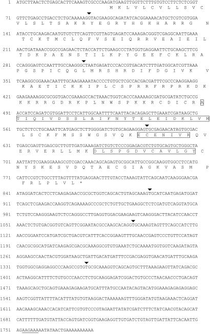
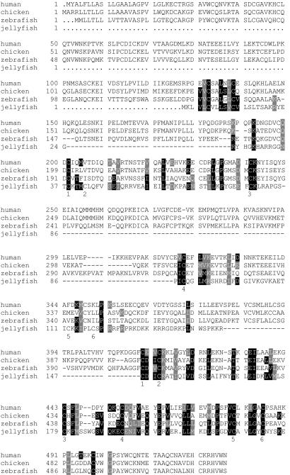
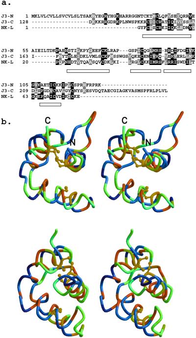
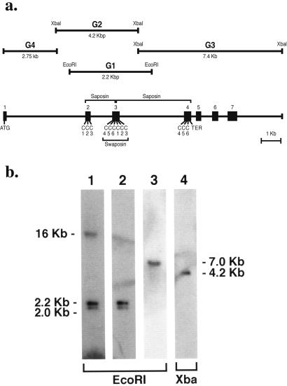
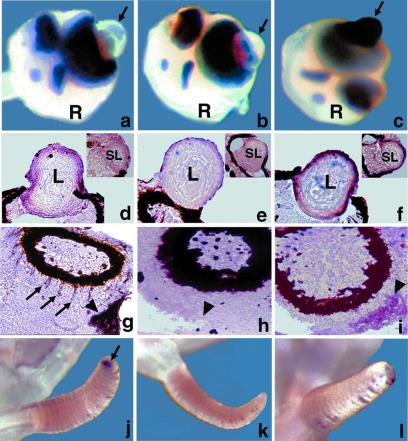
J3-crystallin, one of the three major eye-lens proteins of the cubomedusan jellyfish (Tripedalia cystophora), shows similarity to vertebrate saposins, which are multifunctional proteins that bridge lysosomal hydrolases to lipids and activate enzyme activity. Sequence alignment of deduced J3-crystallin indicates two saposin-like motifs arranged in tandem, each containing six cysteines characteristic of this protein family. The J3-crystallin cDNA encodes a putative precursor analogous to vertebrate prosaposins. The J3-crystallin gene has seven exons, with exons 2-4 encoding the protein. Exon 3 encodes a circularly permutated saposin motif, called a swaposin, found in plant aspartic proteases. J3-crystallin RNA was found in the cubomedusan lens, statocyst, in bands radiating from the pigmented region of the ocellus, in the tentacle tip by in situ hybridization, and in the embryo and larva by reverse transcription-PCR. Our data suggest a crystallin role for the multifunctional saposin protein family in the jellyfish lens. This finding extends the gene sharing evolutionary strategy for lens crystallins to the cnidarians and indicates that the putative primordial saposin/swaposin J3-crystallin reflects both the chaperone and enzyme connections of the vertebrate crystallins.


Figures

Figure 1
Figure 1
cDNA and deduced protein sequences of J3-crystallin cDNA. The arrowheads signify positions of introns (see Fig. 4). The boxed amino acid sequences match the tryptic peptide sequences obtained from the purified protein (JFS-NT12: ECENIVR; JFS3-NT37: DLSPGDV-AVLGY; JFS3-NT53: ATIQIVDSSLAIF-YTELE-DKLVM). The putative polyadenylation signal is underlined.
Figure 2
Figure 2
Sequence comparison of J3-crystallin with human, chicken, and zebrafish prosaposin. Prosaposin amino acids that are identical to those of J3-crystallin in the alignment are highlighted in black; prosaposin residues that are similar to those in J3-crystallin are highlighted in gray. The six characteristic cysteines identifying the two putative saposin motifs in J3-crystallin are indicated under the J3-crystallin sequence. The accession numbers of the human, chicken, and zebrafish prosaposins are given in Methods.
Figure 3
Figure 3
Evidence for two putative saposin motifs within J3-crystallin and their predicted structure based on comparisons with NK-lysin. (a) Multiple sequence alignment of N- and C-terminal parts of J3-crystallin and NK-lysin. Sequence identities and similarities are shaded black and gray, respectively. Helices in the NK-lysin structure (PDB ID code 1NKL) are represented as rectangles. The alignment introduces no insertions or deletions within these helices. J3-N has conserved five of the six cysteines that are disulfide bonded in NK-lysin; the first one of these in J3-crystallin (Csy-38) precedes that in NK-lysin by one residue. All six cysteines of the putative saposin motif are conserved in J3-C. (b) Wall-eyed view of models of J3-N (Upper) and J3-C (Lower) portions of J3-crystallin based on the 1NKL template structure. All disulfide bonds are conserved. The α-carbon traces are colored according to hydropathy (37) with hydrophobic and hydrophilic residues at the red and blue ends of the spectrum, respectively. The lack of hydrophilic residues in the modeled interiors is evident.
Figure 4
Figure 4
Gene structure and genomic Southern blot analysis of J3-crystallin. (a) The J3-crystallin gene was cloned as follows. Five identical 2.2-kbp genomic fragments (G1) were obtained from an EcoRI genomic library by using J3-crystallin cDNA as a probe. Next, a 4.7-kbp clone (G2) was obtained from a XbaI genomic library by screening with G1. Partial sequencing and PCR with primers derived from the J3-crystallin cDNA were used to identify part of exon 2 and all of exon 3 in G1 and exons 2 and 3 in G2. Exons 4–7 were identified on a 7.4-kbp clone (G3) that was obtained from the XbaI library by screening with three deoxyoligonucleotides (5′-gagtgcgagaacatagtgcga-3′; 5′-ctgtaattattcgaaggaaag-3′; 5′ctgtgcagtgctgggctact-3′) derived further downstream on the J3-crystallin cDNA. The 5′ end of the J3-crystallin gene was determined by cloning a PCR product using primers with EcoRI linkers on their ends. The 5′ primer was derived from the beginning of the cDNA and the 3′ primer was derived from the 5′ end of G2. The resulting clone (G4) contained exon 1 at its 5′ end. (b) Southern blot of genomic J3-crystallin sequences. Lane 1, J3-crystallin cDNA probe; lanes 2 and 4, G1 probe; lane 3, G4 probe.
Figure 5
Figure 5
J3 and J1A-crystallin gene expression in adult jellyfish rhopalia and tentacles. In situ hybridization on whole jellyfish (ac, jl) and cryosections (di). J3-crystallin (a, d, g, j) and J1A-crystallin (c, f, i, l) antisense riboprobes; J3-crystallin sense riboprobe (b, e, h, k). R, rhopalium; L, lens of large ocellus; SL, lens of small ocellus. (a, b, c) Arrows point to the surface of the lens of the large ocellus. (g, h, i) Arrowheads point to the tissue surrounding the lumen of the statocyst. (g) Arrows point to the staining in the outer aspect of the pigmented region of the ocellus. (j) Arrow points to the tip of a tentacle.

References

    1. Wistow G, Piatigorsky J. Annu Rev Biochem. 1988;57:479–504. - PubMed
    1. de Jong W W, Hendriks W, Mulders J W, Bloemendal H. Trends Biochem Sci. 1989;14:365–368. - PubMed
    1. Ingolia T D, Craig E A. Proc Natl Acad Sci USA. 1982;79:2360–2364. - PMC - PubMed
    1. Klemenz R, Frohli E, Steiger R H, Schafer R, Aoyama A. Proc Natl Acad Sci USA. 1991;88:3652–3656. - PMC - PubMed
    1. Horwitz J. Proc Natl Acad Sci USA. 1992;89:10449–10453. - PMC - PubMed

Associated data

LinkOut - more resources