. 2001 Oct 23;98(22):12706-11.
doi: 10.1073/pnas.221272198.

Mycobacterium tuberculosis signal transduction system required for persistent infections

Affiliations

Mycobacterium tuberculosis signal transduction system required for persistent infections

T C Zahrt et al. Proc Natl Acad Sci U S A. .

Abstract

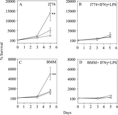
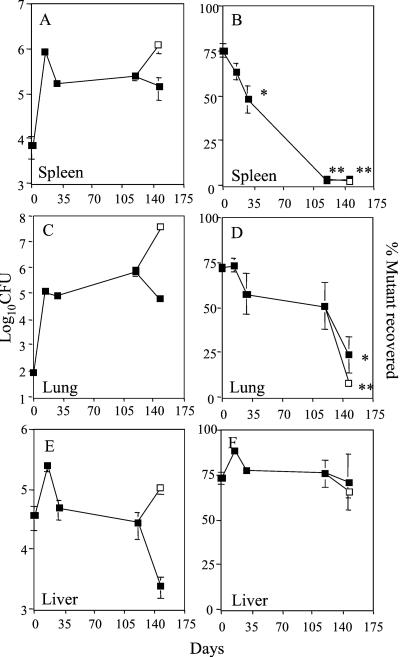
It is estimated that nearly 2 billion people currently suffer from latent Mycobacterium tuberculosis infection. Although the key front-line antituberculosis drugs are effective in treating individuals with acute tuberculosis, these drugs are ineffective in eliminating M. tuberculosis during the persistent stages of latent infection. Consequently, therapeutics that directly target persistent bacilli are urgently needed. We have conducted a global analysis on a group of regulatory determinants that may play a role in M. tuberculosis virulence, and identified a two-component response regulator whose expression is required for entrance into and maintenance of persistent infection. Inactivation of this response regulator, Rv0981 (termed here mprA for mycobacterial persistence regulator), affected M. tuberculosis H37Rv growth in vivo in an organ- and infection stage-specific fashion. These results indicate that two-component systems are important for adaptation of the tubercle bacillus during stages of persistent infection.


Figures

Figure 1
Figure 1
Conservation of M. tuberculosis H37Rv two-component signal transduction systems. Homologs of these genes in other mycobacterial species were: +, present with conserved genetic arrangement; ✻, present with differences in genetic arrangement; or −, absent from the genomes of M. avium (Ma), M. bovis (Mb), M. leprae (Ml), M. smegmatis (Ms), and M. tuberculosis CSU#91 (Mt). When indicated (●), promoter fragments from response regulators were fused to the promoterless gfp gene. Note that not all response regulator promoters could be fused to gfp because of paired arrangements with sensor kinase genes.
Figure 2
Figure 2
In vivo expression of M. tuberculosis response regulators in macrophages. Response regulator promoter fusions were transformed into M. tuberculosis H37Rv or M. bovis bacillus Calmette–Guérin Pasteur and analyzed for expression during growth in macrophages by epifluorescence microscopy. Semiquantitative PCR was used to analyze differences in reporter plasmid copy number between strains. M. tuberculosis response regulator fusions not shown exhibited no detectable fluorescence in M. bovis bacillus Calmette–Guérin or in M. tuberculosis during growth in vitro or during growth in macrophages. +, 0–100 relative fluorescence units (RFU); ++, 100–225 RFU; +++, 225–450 RFU.
Figure 3
Figure 3
Epifluorescence microscopy of macrophage-grown M. tuberculosis H37Rv or M. bovis bacillus Calmette–Guérin representative response regulator promoter fusions from class I (AC), class III (DF), class IV (GI), or the hsp60-gfp control (JL) are shown. Mean fluorescence intensity was determined from M. bovis bacillus Calmette–Guérin-infected J774 monolayers (A, D, G, and J), M. tuberculosis H37Rv-infected J774 monolayers (B, E, H, and K), or M. tuberculosis H37Rv-infected human peripheral blood monocyte-derived macrophages (C, F, I, and L). Insets show intracellular bacilli stained with rhodamine-auramine.
Figure 4
Figure 4
Survival of M. tuberculosis strains growing in macrophages in vitro. Wild-type H37Rv (squares), the Rv0981∷Kmr mutant (diamonds), or Rv0981∷Kmr [attB∷pTZ215] (circles) were used to infect J774 macrophages (J774; A and B) and murine bone marrow-derived macrophages (BMM; C and D). When indicated, macrophages were activated with IFN-γ and LPS 24 h before and during the course of infection (B and D). Bacterial survival has been normalized to the 2-h time point and set to 100%. No growth of M. tuberculosis was observed in tissue culture medium alone. Values represent the mean and standard error of at least three independent experiments. Statistical significance for Rv0981∷Kmr relative to H37Rv or Rv0981∷Kmr [attB∷pTZ215] at each time point: ✻✻, P < 0.01 (ANOVA).
Figure 5
Figure 5
Response regulator Rv0981 (mprA) is required for persistent M. tuberculosis infection. Growth competition experiments were performed between wild-type M. tuberculosis H37Rv and the Rv0981∷Kmr mutant. At various times after infection, colony-forming units (CFU) were determined in the spleen (A), lung (C), and liver (E) of infected mice. Resulting colonies were picked and patched onto selective medium to determine the fraction of Rv0981∷Kmr mutant bacilli recovered (B, D, and F). A subset of infected mice (open squares) received a s.c. injection of hydrocortisone 10 and 5 days before the 147-day time point. Values represent the mean and standard error of total colony-forming units or percent of mutant bacteria recovered from organs. Statistical significance compared percent of mutant recovered at 14, 28, 119, and 147 days after infection relative to the percent of mutant recovered 1 day after infection: ✻, P < 0.05; ✻✻, P < 0.01 (ANOVA).

References

    1. Dye C, Scheele S, Dolin P, Pathania V, Raviglione M C. J Am Med Assoc. 1999;282:677–686. - PubMed
    1. Manabe Y C, Bishai W R. Nat Med. 2000;6:1327–1329. - PubMed
    1. Camacho L R, Ensergueix D, Perez E, Gicquel B, Guilhot C. Mol Microbiol. 1999;34:257–267. - PubMed
    1. Berthet F X, Lagranderie M, Gounon P, Laurent-Winter C, Ensergueix D, Chavarot P, Thouron F, Maranghi E, Pelicic V, Portnoi D, et al. Science. 1998;282:759–762. - PubMed
    1. Li Z, Kelley C, Collins F, Rouse D, Morris S. J Infect Dis. 1998;177:1030–1035. - PubMed

Publication types

Substances