. 2001 Oct 23;98(22):12730-5.
doi: 10.1073/pnas.231471298.

Transient uptake of serotonin by newborn olfactory projection neurons

Affiliations

Transient uptake of serotonin by newborn olfactory projection neurons

B S Beltz et al. Proc Natl Acad Sci U S A. .

Abstract

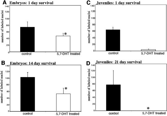
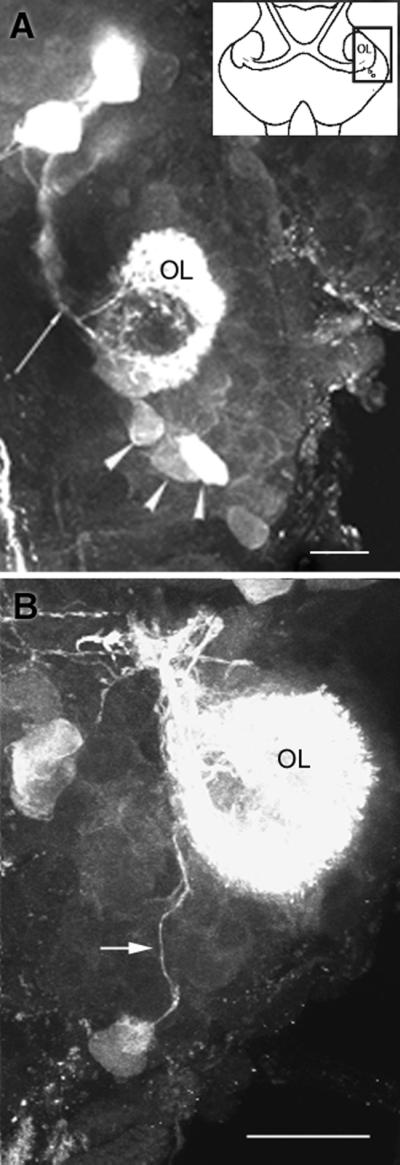
A life-long turnover of sensory and interneuronal populations has been documented in the olfactory pathways of both vertebrates and invertebrates, creating a situation where the axons of new afferent and interneuronal populations must insert into a highly specialized glomerular neuropil. A dense serotonergic innervation of the primary olfactory processing areas where these neurons synapse also is a consistent feature across species. Prior studies in lobsters have shown that serotonin promotes the branching of olfactory projection neurons. This paper presents evidence that serotonin also regulates the proliferation and survival of projection neurons in lobsters, and that the serotonergic effects are associated with a transient uptake of serotonin into newborn neurons.


Figures

Figure 1
Figure 1
The olfactory pathway in the brain of Homarus americanus. (A) Confocal image of the OLs and ALs in a hemi-brain of an embryonic lobster stained with an antibody against Drosophila synapsin (E. Buchner, Würzburg, Germany). Tissues were processed by standard methods (22). The diverging arrows mark the path of the primary neurites of the olfactory projection neurons (somata located in cluster 10) that innervate the OL and AL. These processes form the OGT (converging arrows) that passes through the OGTN (*) en route to the lateral protocerebrum. (B) Schematic diagram illustrating the position of the neuropils that are altered by serotonin depletion during embryonic development: OL, AL, and OGTN. (Left Inset) The area of synapsin labeling shown in A. (Right Inset) The area of serotonin labeling shown in C. (C) Serotonin immunostaining (for method, see ref. 21) of the DGN, which innervates the ipsilateral OL, AL, and OGTN (not shown). The intense labeling of the OL and AL is due to the massive DGN projection into these areas (16, 22). (Bars: A = 20 μm; C = 200 μm.) [A is reprinted with permission from B. S. Beltz and colleagues (23) (Copyright 2000, Society for Neuroscience).]
Figure 2
Figure 2
Persistent cell proliferation in cluster 10, which contains the somata of the olfactory projection neurons, in juvenile and adult brains. (A) Newly born projection neurons are labeled in 0.5-year-old juveniles by injecting BrdUrd 3 weeks before killing and with a second pulse on the day of killing. The dividing cells labeled by the 24-h pulse are located in the proliferation zone (double arrowheads), whereas the cells labeled by the injection 3 weeks before killing have migrated anterolaterally (single arrowhead; ref. 5). (B) BrdUrd injection of adult lobsters followed by killing 6 weeks later demonstrates a band of labeled cells in cluster 10 (arrowhead) that has moved anterolaterally from their origin at the posteromedial margin of the cluster (5). (C) When BrdUrd was injected into adult lobsters at 6 months and again at 24 h before killing, nuclei are labeled posteromedially in the proliferation zone (double arrowheads), whereas individual labeled cells have dispersed among the cluster 10 neurons. (D) Higher magnification of labeled cells outlined in C. (Bars: AC = 50 μm; D = 20 μm.) [A and B are reprinted with permission from B. S. Beltz and colleagues (5) (Copyright 1999, Society for Neuroscience).]
Figure 3
Figure 3
A comparison of the levels of proliferation (A and C) and survival (B and D) of BrdUrd-labeled cells in cluster 10 of control and 5,7-DHT-treated (experimental) embryonic and juvenile lobsters. The numbers of BrdUrd-labeled profiles were counted and compared with two-tailed t tests. (A, B, and D) *, statistically significant differences (P < 0.0001) between experimental and control groups. Statistical tests were not valid for C because of the low control–sample size. The numbers of labeled cells are represented as means ± SD. (A) To examine the effects of serotonin depletion on neuronal proliferation during embryogenesis, control (n = 10 cell clusters) and 5,7-DHT-treated (n = 10 cell clusters) embryos were exposed to BrdUrd for 2 h and killed after 24 h. The number of BrdUrd-labeled cells in cluster 10 in 5,7-DHT-treated embryos was found to be on average 35% smaller than that observed in control embryos. (B) To study the longer term effects of serotonin depletion, control (n = 10 cell clusters) and 5,7-DHT-treated (n = 10 cell clusters) embryos were killed 2 weeks after a 2-h period of exposure to BrdUrd. By using this protocol, the number of BrdUrd-labeled cells observed in 5,7-DHT-treated embryos was found to be 52% smaller than the number of labeled cells in control embryos. (C and D) Serotonin depletion in juvenile lobsters was found to reduce the numbers of labeled profiles when assessed 24 h (C) (control, n = 2 cell clusters; 5,7-DHT-treated, n = 4 cell clusters) and 3 weeks (D) (control, n = 4 cell clusters; 5,7-DHT-treated, n = 4 cell clusters) after BrdUrd injection. (C) Whereas an average of 78 neurons was labeled in each cluster 10 of control lobsters 24 h after BrdUrd injection, an average of only 4 neurons was labeled in serotonin-depleted lobsters. (D) No labeled neurons were observed in the brains of 5,7-DHT-treated lobsters after a 3-week survival time after BrdUrd injection.
Figure 4
Figure 4
Confocal images of the brains of juvenile lobsters labeled for BrdUrd (red) and serotonin (green) immunoreactivities. (A) Lobsters were killed 24 h after BrdUrd injection, and dissected brains were incubated in serotonin, rinsed, fixed, and processed by standard methods. BrdUrd-labeled nuclei are found at the anterior margin of the proliferation zone in cluster 10. Serotonin labeling is seen in the AL and in a row of six labeled cells (arrowheads) that lie between the BrdUrd profiles and the AL; one of these cells is double-labeled (yellow arrowhead). The positions of the serotonin-labeled cells relative to the BrdUrd-labeled nuclei suggest that these cells were born after the BrdUrd-labeled neurons that have migrated anterolaterally during the 24-h survival time (n = 4 cell clusters). (B) After a 6-h survival time after BrdUrd injection, labeled nuclei are found in the proliferation zone immediately adjacent to the AL. Serotonin antibodies label a group of fine fibers (arrow) that terminate blindly at the proliferation zone. These fibers have been traced back to the DGN in adult, juvenile, and larval brains. In embryos, the narrow region to which these fibers project is obscured by the intense labeling for serotonin in newborn cells, and, therefore, it has not been possible to trace the fibers to the DGN. (Bars: A = 100 μm; B = 50 μm.)
Figure 5
Figure 5
Confocal images of serotonin labeling in embryonic brains. (A) In lobsters at 35% of embryonic development, projection neurons with somata located at the posterior margin of cluster 10 (arrowheads) take up serotonin transiently (n = 12 cell clusters). Unlabeled projection neuron somata can be seen just anterior and lateral to the stained cells. The AL has not emerged at this time in development. Fibers from local interneurons (arrow), including the DGN, also label for serotonin. (B) The axons of serotonin-labeled cluster 10 neurons can be traced to the embryonic OL. (Bars = 20 μm.)

Similar articles

Cited by

References

    1. Graziadei P P C, Monti Graziadei G A. Neuroscience. 1986;19:1025–1035. - PubMed
    1. Harrison P J, Cate H S, Swanson E S, Derby C D. J Neurobiol. 2001;47:51–66. - PubMed
    1. Lois C, Alvarez-Buylla A. Science. 1994;264:1145–1148. - PubMed
    1. Sandeman R, Clarke D, Sandeman D, Manly M. J Neurosci. 1998;18:6195–6206. - PMC - PubMed
    1. Harzsch S, Miller J, Benton J, Beltz B S. J Neurosci. 1999;19:3472–3485. - PMC - PubMed

Publication types

LinkOut - more resources