. 2001 Nov;67(11):5273-84.
doi: 10.1128/AEM.67.11.5273-5284.2001.

In situ characterization of Nitrospira-like nitrite-oxidizing bacteria active in wastewater treatment plants

Affiliations

In situ characterization of Nitrospira-like nitrite-oxidizing bacteria active in wastewater treatment plants

H Daims et al. Appl Environ Microbiol. 2001 Nov.

Abstract

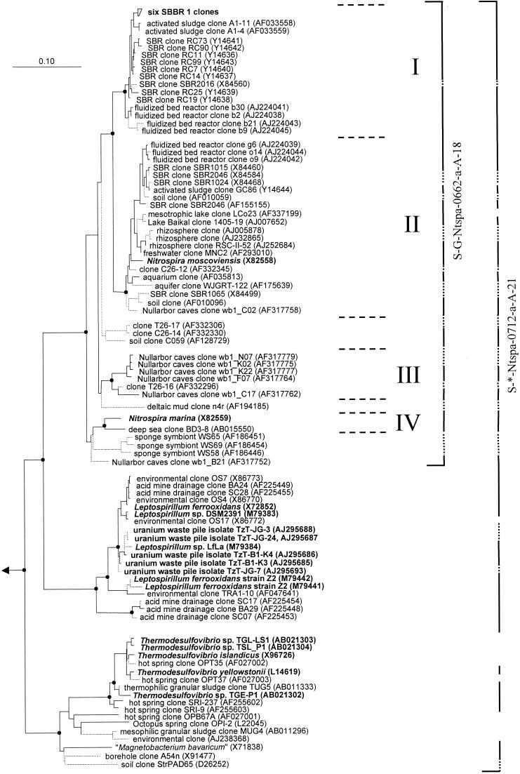
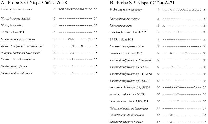
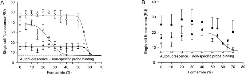
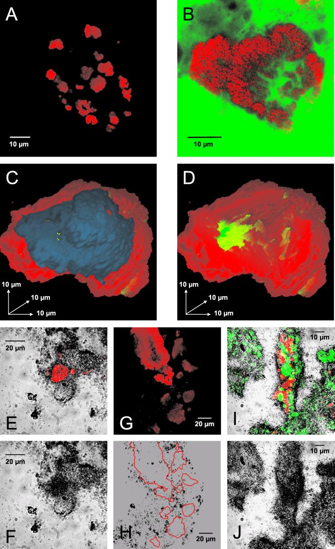
Uncultivated Nitrospira-like bacteria in different biofilm and activated-sludge samples were investigated by cultivation-independent molecular approaches. Initially, the phylogenetic affiliation of Nitrospira-like bacteria in a nitrifying biofilm was determined by 16S rRNA gene sequence analysis. Subsequently, a phylogenetic consensus tree of the Nitrospira phylum including all publicly available sequences was constructed. This analysis revealed that the genus Nitrospira consists of at least four distinct sublineages. Based on these data, two 16S rRNA-directed oligonucleotide probes specific for the phylum and genus Nitrospira, respectively, were developed and evaluated for suitability for fluorescence in situ hybridization (FISH). The probes were used to investigate the in situ architecture of cell aggregates of Nitrospira-like nitrite oxidizers in wastewater treatment plants by FISH, confocal laser scanning microscopy, and computer-aided three-dimensional visualization. Cavities and a network of cell-free channels inside the Nitrospira microcolonies were detected that were water permeable, as demonstrated by fluorescein staining. The uptake of different carbon sources by Nitrospira-like bacteria within their natural habitat under different incubation conditions was studied by combined FISH and microautoradiography. Under aerobic conditions, the Nitrospira-like bacteria in bioreactor samples took up inorganic carbon (as HCO(3)(-) or as CO(2)) and pyruvate but not acetate, butyrate, and propionate, suggesting that these bacteria can grow mixotrophically in the presence of pyruvate. In contrast, no uptake by the Nitrospira-like bacteria of any of the carbon sources tested was observed under anoxic or anaerobic conditions.


Figures

FIG. 1
FIG. 1
Phylogenetic tree of the phylum Nitrospira based on comparative analysis of 16S rRNA sequences. The basic tree topology was determined by maximum-likelihood analysis of all sequences longer than 1,300 nucleotides. Shorter sequences were successively added by use of the ARB_PARSIMONY module of the ARB program without changing the overall tree topology. Branches leading to sequences shorter than 1,000 nucleotides are dotted to point out that the exact affiliation of these sequences cannot be determined. Black spots on tree nodes symbolize high-parsimony bootstrap support above 90% based on 100 iterations. The scale bar indicates 0.1 estimated change per nucleotide. Sequences of Nitrospira-like bacteria retrieved in this study from reactor SBBR 1 and sequences that belong to isolated strains are in boldface. The four sublineages of the genus Nitrospira are delimited by horizontal dashed lines and numbered I to IV. The brackets illustrate the coverage of the 16S rRNA-targeted oligonucleotide probes developed in this study. Dotted bracket segments indicate that the corresponding partial sequences do not include the probe target site. Brackets are interrupted where sequences are not targeted by the respective probe.
FIG. 2
FIG. 2
Target site sequences and corresponding 16S rRNA sequence regions of target and nontarget organisms for probe S-G-Ntspa-0662-a-A-18 (A) and probe S-*-Ntspa-0712-a-A-21 (B). Hyphens represent identical nucleotides. Mismatches between the rRNA sequences of organisms and the probe target site sequence are indicated by capital letters.
FIG. 3
FIG. 3
Probe dissociation profiles of the oligonucleotide probes developed in this study with reference organisms under increasingly stringent hybridization and washing conditions. For each data point, the mean fluorescence intensity of at least 100 cells was determined. Regression curves were calculated by the plotting software based on a sigmoidal curve fit model. Error bars indicate 1 standard deviation. Error bars that are smaller than the marker symbols are not shown. (A) Hybridization of target organism N. moscoviensis with probe S-G-Ntspa-0662-a-A-18 in the presence of Comp-Ntspa-0662 (▴). Hybridization of nontarget bacterium B. stearothermophilus with probe S-G-Ntspa-0662-a-A-18 without (○) and with (Δ) competitor Comp-Ntspa-0662. (B) Hybridization of target organisms N. moscoviensis (●) and L. ferrooxidans (■) with probe S-*-Ntspa-0712-a-A-21 in the presence of competitor Comp-Ntspa-0712. Hybridization of nontarget bacterium D. desulfuricans with probe S-*-Ntspa-0712-a-A-21 without (▵) and with (□) competitor Comp-Ntspa-0712. The regression curve refers to the data points obtained for D. desulfuricans without addition of the competitor. RU, relative units.
FIG. 4
FIG. 4
In situ analyses of Nitrospira-like bacteria within activated sludge and biofilms. (A) Nitrospira cell aggregates detected in activated sludge by FISH with probe S-G-Ntspa-0662-a-A-18 (red). (B) Nitrospira cell aggregate detected in biofilm from reactor SBBR 1 by FISH with probe S-G-Ntspa-0662-a-A-18 (red). All of the Nitrospira colonies detected in SBBR 1 with probe S-G-Ntspa-0662-a-A-18 also hybridized with probe S-*-Ntspa-0712-a-A-21 (data not shown). The biofilm was also stained with fluorescein (green). (C and D) Three-dimensional reconstruction of a Nitrospira cell aggregate from reactor SBBR 1 stained by FISH with probe S-G-Ntspa-0662-a-A-18. Nitrospira cells are red; the blue part of the microcolony in panel C was digitally removed to allow insight into the aggregate (D). Voids within the aggregate are green. (E and F) Uptake of bicarbonate by Nitrospira-like bacteria in biofilm from reactor SBBR 1 under aerobic incubation conditions. (E) Nitrospira cells stained by probe S-G-Ntspa-0662-a-A-18 (red) combined with the micrograph of the radiographic film at the same position. Panel F shows only the film to visualize the MAR signal at the position of the Nitrospira cells. Other MAR signals were caused by CO2-fixing bacteria, which were not detected by the Nitrospira-specific probe. (G and H) No uptake of acetate by Nitrospira-like bacteria in biofilm from reactor SBBR 1 under aerobic incubation conditions. (G) Nitrospira cells stained by probe S-G-Ntspa-0662-a-A-18 (red). Panel H is a micrograph of the radiographic film at the same position. The localization of the Nitrospira microcolonies in panel G is indicated by red borderlines in panel H. (I and J) Uptake of pyruvate by Nitrospira-like bacteria (stained by probe S-G-Ntspa-0662-a-A-18; red) and by ammonia oxidizers (stained by probes NEU and Nso1225; green) in biofilm from reactor SBBR 1 under aerobic incubation conditions. The fluorescence recorded in a stack of images by the CLSM is combined by orthographic projection in panel I. Stacked cells of Nitrospira-like bacteria and ammonia oxidizers appear therefore yellow. The image stack was acquired to ensure that all of the nitrifiers that contributed to the MAR signal would be visible in the final image.
FIG. 5
FIG. 5
Concentrations of NH4-N (●), NO3-N (▪), and NO2-N (▴) in reactor SBBR 1 during a representative reactor operating cycle.

Similar articles

Cited by

References

    1. Alm E W, Oerther D B, Larsen N, Stahl D A, Raskin L. The oligonucleotide probe database. Appl Environ Microbiol. 1996;62:3557–3559. - PMC - PubMed
    1. Amann R I. In situ identification of micro-organisms by whole cell hybridization with rRNA-targeted nucleic acid probes. In: Akkeman A D C, van Elsas J D, de Bruigin F J, editors. Molecular microbial ecology manual. 3.3.6. Dordrecht, The Netherlands: Kluwer Academic Publishers; 1995. pp. 1–15.
    1. Amann R I, Binder B J, Olson R J, Chisholm S W, Devereux R, Stahl D A. Combination of 16S rRNA-targeted oligonucleotide probes with flow cytometry for analyzing mixed microbial populations. Appl Environ Microbiol. 1990;56:1919–1925. - PMC - PubMed
    1. Amann R I, Ludwig W, Schleifer K-H. Phylogenetic identification and in situ detection of individual microbial cells without cultivation. Microbiol Rev. 1995;59:143–169. - PMC - PubMed
    1. Bever J, Stein A, Teichmann H, editors. Weitergehende Abwasserreinigung. Munich, Germany: R. Oldenbourg Verlag; 1995.

Publication types

MeSH terms

Associated data

LinkOut - more resources