

The xenograft antigen in complex with GS-1-B4 lectin: crystallization and preliminary X-ray analysis
- PMID: 11679730
- PMCID: PMC4190837
- DOI: 10.1107/s0907444901012148
The xenograft antigen in complex with GS-1-B4 lectin: crystallization and preliminary X-ray analysis
Abstract
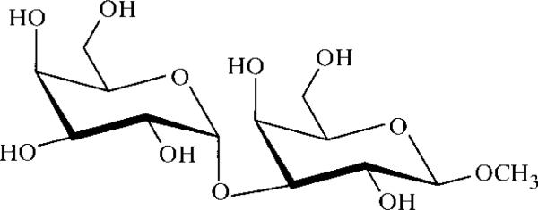
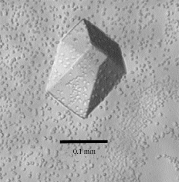
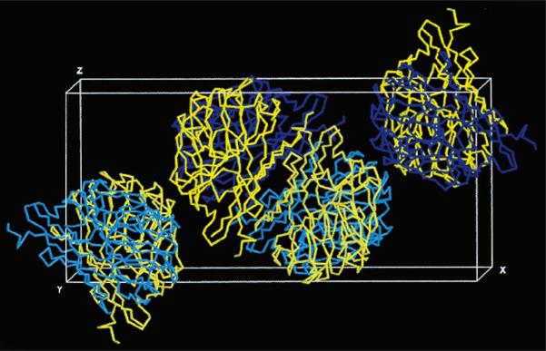
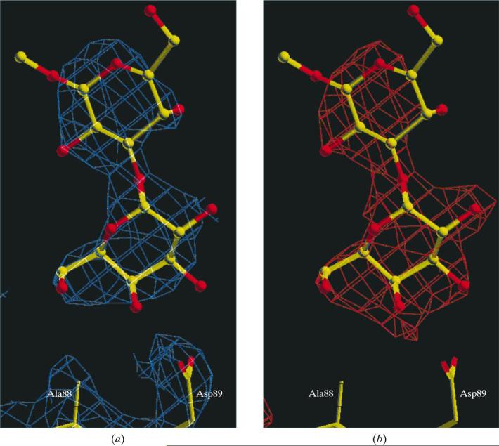
The implantation of animal organs is one approach to overcoming the shortage of human donor organs for medical transplantation. Although readily available, non-primate tissues are subject to hyperacute rejection wherein human anti-Galalpha(1-3)Gal antibodies react with haptens present on the transplanted cells' surfaces. The understanding of this interaction on a molecular level will further the development of a strategy for the prevention of hyperacute rejection in xenotransplantation. The Galalpha(1-3)Gal hapten ('xenograft antigen') has been cocrystallized with the Gal-specific B(4) isolectin of Griffonia simplicifolia lectin-1. Crystals were analyzed by cryocrystallography and were found to diffract to moderately high resolution on a rotating-anode X-ray source. They belong to the P2(1)2(1)2 space group, with unit-cell parameters a = 111.0, b = 51.3, c = 76.9 A, and contain two molecules per asymmetric unit.
Figures

References
-
- Altschul SF, Gish W, Miller W, Myers EW, Lipman DJ. J. Mol. Biol. 1990;215:403–410. - PubMed
-
- Bernstein HJ. RasMol Version 2.7.1.1. 2001 http://www.bernstein-plus-sons.com/software/rasmol/
-
- Brunger AT, Adams PD, Clore GM, DeLano WL, Gros P, Grosse-Kunstleve RW, Jiang JS, Kuszewski J, Nilges M, Pannu NS, Read RJ, Rice LM, Simonson T, Warren GL. Acta Cryst. 1998;D54:905–921. - PubMed
-
- Christie KN, Thomson C. J. Histochem. Cytochem. 1989;37:1303–1304. - PubMed
-
- DeLano WL, Brünger AT. Acta Cryst. 1995;D51:740–748. - PubMed
Publication types
MeSH terms
Substances
Associated data
- Actions
Grants and funding
LinkOut - more resources
Full Text Sources
