. 2001 Oct;104(2):135-41.
doi: 10.1046/j.1365-2567.2001.01289.x.

Size of the population of CD4+ natural killer T cells in the liver is maintained without supply by the thymus during adult life

Affiliations

Size of the population of CD4+ natural killer T cells in the liver is maintained without supply by the thymus during adult life

H Kameyama et al. Immunology. 2001 Oct.

Abstract

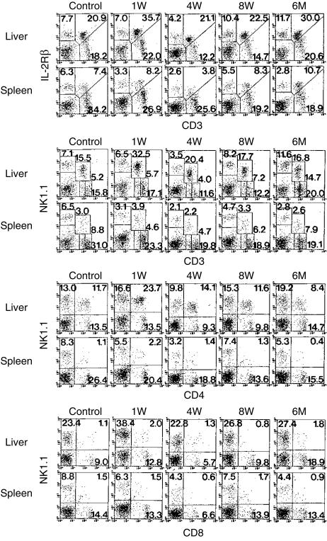
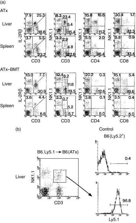
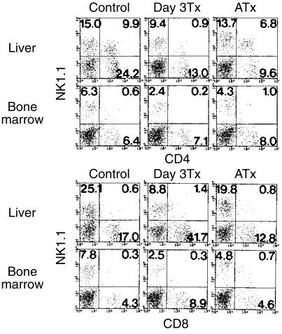
Given that there are few natural killer T (NKT) cells in the liver of athymic nude mice and in neonatally thymectomized mice, it is still controversial whether all NKT cells existing in the liver are supplied by the thymus or if some such cells develop in the liver. To determine whether or not NKT cells are consistently supplied from the thymus during adult life, thymectomy was conducted in mice at the age of 8 weeks. Interestingly, the proportion and number of CD4+ NKT cells increased or remained unchanged in the liver after adult thymectomy and this phenomenon continued for up to 6 months after thymectomy. The administration of alpha-galactosylceramide induced severe cytopenia (due to apoptosis) of CD4+ NKT cells in the liver on day 1, but subsequent expansion of these NKT cells occurred in thymectomized mice similar to the case in normal mice. However, in thymectomized mice given lethal irradiation (9.5 Gy) and subsequent bone marrow transfer, the population of CD4+ NKT cells no longer expanded in the liver, although that of CD8+ NKT cells did. These results suggest that thymic CD4+ NKT cells, or their progenitors, may migrate to the liver at a neonatal stage but are not supplied from the thymus in the adult stage under usual conditions. CD8+ NKT cells can be generated in the liver.


Figures

Figure 1
Figure 1
Phenotypic characterization of lymphocytes in the liver and spleen of mice with adult thymectomy. B6 mice at the age of 8 weeks were thymectomized. Lymphocytes were then obtained from the liver and spleen at the indicated weeks after thymectomy. Two-colour staining for CD3 and IL-2Rβ, that for CD3 and NK1.1, that for CD4 and NK1.1, and that for CD8 and NK1.1 were conducted. Numbers in the figure represent the percentages of fluorescence-positive cells. The data shown here are representative of three experiments.
Figure 2
Figure 2
Administration of α-GalCer to thymectomized mice. Mice were kept for 2 weeks after adult thymectomy and were then intraperitoneally injected with α-GalCer (2 µg/mouse). Two-colour staining for the indicated combinations was conducted on days 1, 3 and 7 after the injection. Numbers in the figure represent the percentages of fluorescence-positive cells. The data shown here are representative of three experiments.
Figure 3
Figure 3
Induction of hepatic injury by administration of α-GalCer in mice with adult thymectomy. Mice were kept for 2 weeks after adult thymectomy and were then intraperitoneally injected with α-GalCer (2 µg/mouse). The data for serum transaminases, GOT and GPT, at each point were produced using four mice.
Figure 4
Figure 4
A comparison of the phenotypic characterization of lymphocytes in the liver between thymectomized mice and thymectomized mice irradiated lethally and subjected to BMT. (a) ATx and ATx-BMT, (b) ATx-BMT (Ly5.1 origin). Lymphocytes were obtained from the liver of thymectomized mice lethally irradiated and subjected to BMT at 2 months after such treatments. Age-matched control (thymectomized) mice were also used. Two-colour staining for the indicated combinations was conducted. In (b), BMT of B6.Ly5.1 origin was conducted to determine the origin of newly generated CD8+ NKT cells in ATxBMT mice (B6.Ly5.2+). Three-colour staining for CD8, NK1.1, and Ly5.1 was conducted. The data shown here are representative of three experiments.
Figure 5
Figure 5
Size of the population of CD4+ and CD8+ NKT cells in the liver and bone marrow after neonatal or adult thymectomy. Neonatal thymectomy was performed by day 3 after birth (Day 3 Tx) while adult thymectomy was done at the age of 8 weeks (ATx). All mice, including untreated B6 mice, were used at the age of 6 months. Two-colour staining for the indicated combinations was conducted. The data shown here are representative of three experiments.

References

    1. Bendelac A. Mouse NK1+ T cells. Curr Opin Immunol. 1995;7:367–74. - PubMed
    1. MacDonald HR. NK1.1+ T cells receptor-α/β+ cells: new clues to their origin, specificity, and function. J Exp Med. 1995;182:633–8. - PMC - PubMed
    1. Bix M, Locksley RM. Natural T cells: Cells that co-express NKRP-1 and TCR. J Immunol. 1995;155:1020–2. - PubMed
    1. Vicari AP, Zlotnik A. Mouse NK1.1+ T cells: a new family of T cells. Immunol Today. 1996;17:71–6. - PubMed
    1. Abo T, Kawamura T, Watanabe H. Physiological responses of extrathymic T cells in the liver. Immunol Rev. 2000;174:135–49. - PubMed

Publication types

Substances