. 2001 Oct;104(2):215-25.
doi: 10.1046/j.1365-2567.2001.01287.x.

Tissue distribution of products of the mouse decay-accelerating factor (DAF) genes. Exploitation of a Daf1 knock-out mouse and site-specific monoclonal antibodies

Affiliations

Tissue distribution of products of the mouse decay-accelerating factor (DAF) genes. Exploitation of a Daf1 knock-out mouse and site-specific monoclonal antibodies

F Lin et al. Immunology. 2001 Oct.

Abstract

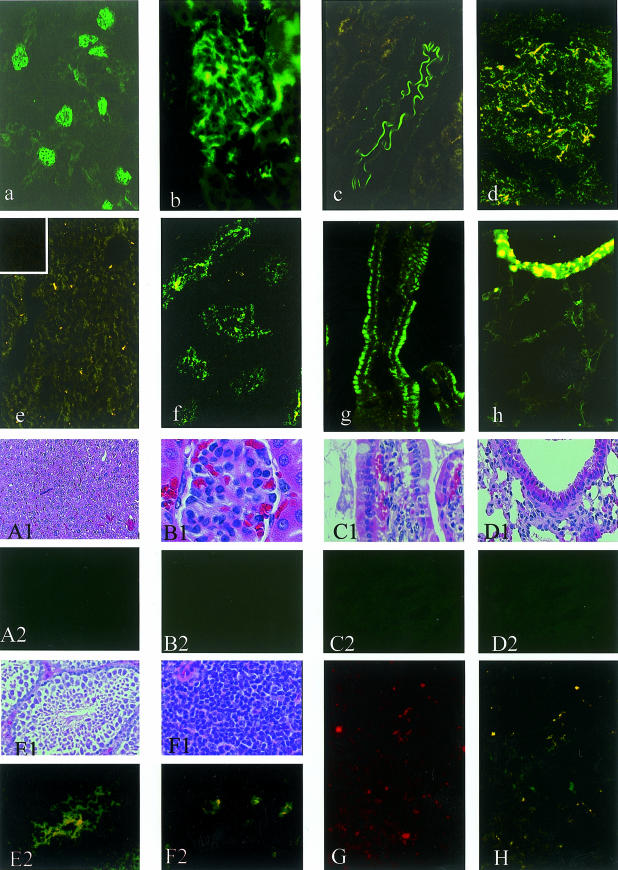
Decay-accelerating factor (DAF) is a membrane regulator of C3 activation that protects self cells from autologous complement attack. In humans, DAF is uniformly expressed as a glycosylphosphatidylinositol (GPI)-anchored molecule. In mice, both GPI-anchored and transmembrane-anchored DAF proteins are produced, each of which can be derived from two different genes (Daf1 and Daf2). In this report, we describe a Daf1 gene knock-out mouse arising as the first product of a strategy for targeting one or both Daf genes. As part of the work, we characterize recently described monoclonal antibodies against murine DAF protein using deletion mutants synthesized in yeast, and then employ the monoclonal antibodies in conjunction with wild-type and the Daf1 knock-out mice to determine the tissue distribution of the mouse Daf1 and Daf2 gene products. To enhance the immunohistochemical detection of murine DAF protein, we utilized the sensitive tyramide fluorescence method. In wild-type mice, we found strong DAF labelling of glomeruli, airway and gut epithelium, the spleen, vascular endothelium throughout all tissues, and seminiferous tubules of the testis. In Daf1 knock-out mice, DAF labelling was ablated in most tissues, but strong labelling of the testis and splenic dendritic cells remained. In both sites, reverse transcription-polymerase chain reaction analyses identified both GPI and transmembrane forms of Daf2 gene-derived protein. The results have relevance for studies of in vivo murine DAF function and of murine DAF structure.


Figures

Figure 1
Figure 1
Targeting strategy for inactivation of the mouse Daf genes. The tandem Daf1 and Daf2 genes are shown diagrammatically (not drawn to scale). The black-filled boxes represent exons and the open boxes represent selection marker genes as marked. As the figure shows, the pDAFup construct integrates to the Daf1 gene first to introduce a loxP site and a TK gene within exon 3, then the pDAFdown construct may integrate into its homologous region in either the Daf1 or Daf2 gene together with another loxP site and TK gene. Upon recombination induced by Cre recombinase, the DNA fragment flanked by the two loxP sites is deleted together with the two TK genes to generate either Daf1 or Daf2 knock-out ES cells.
Figure 2
Figure 2
PCR analysis of recombined ES cells. (a1) PCR with Neo and Hph-specific primers P3 and P4 yielded a ∼500-bp fragment verifying that the two markers were brought together by Cre/loxP recombination. M, 1 kb ladder; C, PCR with the wild-type DNA as template; K/O, recombined DNA as template. (a2) PCR with primers P5, P6 (Daf1-specific) and P7 (Daf2-specific) showing that the pDAFdown construct integrated into exon 5 of the Daf1 gene and that the Daf1 gene thus was selectively inactivated. (a3) RT-PCR with primers P8 and P10 of the Daf1 mRNA product in the Daf1−/− mice. A truncated product corresponding to sequence for CCP1,4 is seen. (b) A diagram of the mouse Daf1 and Daf2 genes is shown. B, BamHI; E, EcoRI; and S, SacI. The position of the 1·5-kb SacI probe is indicated and the hybridized EcoRI and BamHI fragments are shown by the brackets. (c) Southern blot analyses of EcoRI- and BamHI-digested genomic DNA from parental and K/O mice. DNA from wild-type, heterozygous (Daf1+/−) and homozygous (Daf1−/−) knock-out mice were hybridized with the SacI fragment of the Daf1 gene (panel B). The pattern corresponded to the expected deletion from Daf1 exon 3 to exon 5. The high Mr band corresponds to the homologous EcoRI fragment in the Daf2 gene.
Figure 3
Figure 3
Functional analyses of Daf1-derived CCP1,4 protein. (a) and (b), EshA were incubated with mouse serum in the presence of the purified Daf1 CCP1,4 product, purified native CCP1-4 protein, or BSA. (a) FACS analyses showing that C3b deposition on EshA was unaffected by the CCP1,4 product. (b) Dose–response curve of the relative inhibitory activities of the DAF1,4 product and native DAF1-4 as compared to BSA on C3b deposition on EshA. (c) CD97-transfected COS cells were incubated with erythrocytes from wild-type and the Daf1 gene-targeted mice. The number of adherent erythrocytes per high powered field (∼50 COS cell transfectants) was scored.
Figure 4
Figure 4
(a) FACS analyses of erythrocytes from wild-type and Daf1−/− mice using 2C6 rat anti-murine DAF mAb. (b) Similar analyses of splenic suspension cells from wild-type and Daf1−/− mice. The unfilled peaks show the staining with isotype-matched non-relevant control mAb. (c) FACS analyses of autologous C3 deposition in vivo on erythrocytes from Daf1−/− and wild-type littermates. The bars show the average values (P < 0·005).
Figure 5
Figure 5
Immunostaining of DAF in wild-type mice and Daf1−/− mice. All tissues were stained with 2C6 mAb. In sections from wild-type mice (a through h), the kidney at ×100 magnification (a), a glomerulus at ×400 magnification (b) and a renal artery at ×400 magnification (c) showed intense labelling. The spleen at ×100 magnification (d) showed heavy staining of the white pulp, with weaker focal staining of the red pulp. The liver at ×100 magnification (e) revealed moderate endothelial staining in larger blood vessels, but weak labelling of sinusoidal endothelium (inset in corner shows negative control). Epithelial staining was detected in the testis at ×100 magnification (f), the villi of the small intestine at ×200 magnification (g) and the bronchi at ×400 magnification (h). In corresponding but not identical sections from Daf1−/− mice stained with H&E (A1–F1) and with 2C6 mAb (A2–F2), the liver at ×100 (A1, A2), a glomerulus at ×400 (B1, B2), intestinal villi at ×200 (C1, C2) and bronchi at ×400 (D1, D2) were completely negative. In contrast, intense staining was seen in the testis at ×400 magnification (E1, E2) and in dendritic cells in the spleen at ×400 magnification (F1, F2). Staining of the other duplicate spleen sections from Daf1−/− mice with 2C6 mAb at ×100 (G) and with CD11c mAb at ×100 (H) showed an identical staining pattern of dendritic cell labelling.
Figure 6
Figure 6
(a) RT-PCR analysis of Daf mRNA expression in different tissues in wild-type and Daf1−/− mice. Parallel amplification of β-actin was included as a control. M, 1 kb ladder; 1, Daf1; 2, Daf2; A, β-actin. (b) RT-PCR analyses of Daf2 mRNA in spleen and testis of Daf1−/− mice using GPI-and TM-specific primers P13 and P14 in conjunction with P9. In both sites, the TM form of the transcript is seen. TM, TM-DAF; GPI, GPI-DAF.

References

    1. Nussenzweig V. Inhibition of complement activation on the surface of cells after incorporation of decay-accelerating factor (DAF) into their membranes. J Exp Med. 1984;160:1558–78. - PMC - PubMed
    1. Nicholson-Weller A, Burge J, Fearon DT, Weller PF, Austen KF. Isolation of a human erythrocyte membrane glycoprotein with decay-accelerating activity for C3 convertases of the complement system. J Immunol. 1982;129:184–9. - PubMed
    1. Pangburn MK, Schreiber RD, Muller-Eberhard HJ. Deficiency of an erythrocyte membrane protein with complement regulatory activity in paroxysmal nocturnal hemoglobinuria. Proc Natl Acad Sci USA. 1983;80:5430–4. - PMC - PubMed
    1. Fujita T, Inoue T, Ogawa K, Iida K, Tamura N. The mechanism of action of decay-accelerating factor (DAF). DAF inhibits the assembly of C3 convertases by dissociated C2a and Bb. J Exp Med. 1987;166:1221–8. - PMC - PubMed
    1. Lublin DM, Lemons RS, Beau MML, Holers VM, Tykocinski ML, Medof ME, Atkinson JP. The gene encoding decay-accelerating factor (DAF) is located in the complement regulatory locus on the long arm of chromosome 1. J Exp Med. 1987;165:1731–6. - PMC - PubMed

Publication types