. 2001 Oct 29;155(3):339-54.
doi: 10.1083/jcb.200108007. Epub 2001 Oct 29.

Novel vertebrate nucleoporins Nup133 and Nup160 play a role in mRNA export

Affiliations

Novel vertebrate nucleoporins Nup133 and Nup160 play a role in mRNA export

S Vasu et al. J Cell Biol. .

Abstract

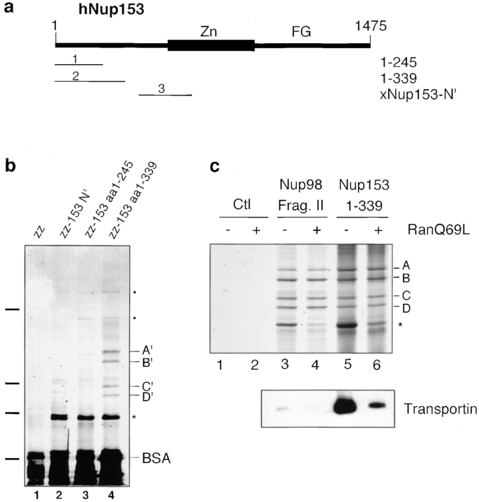
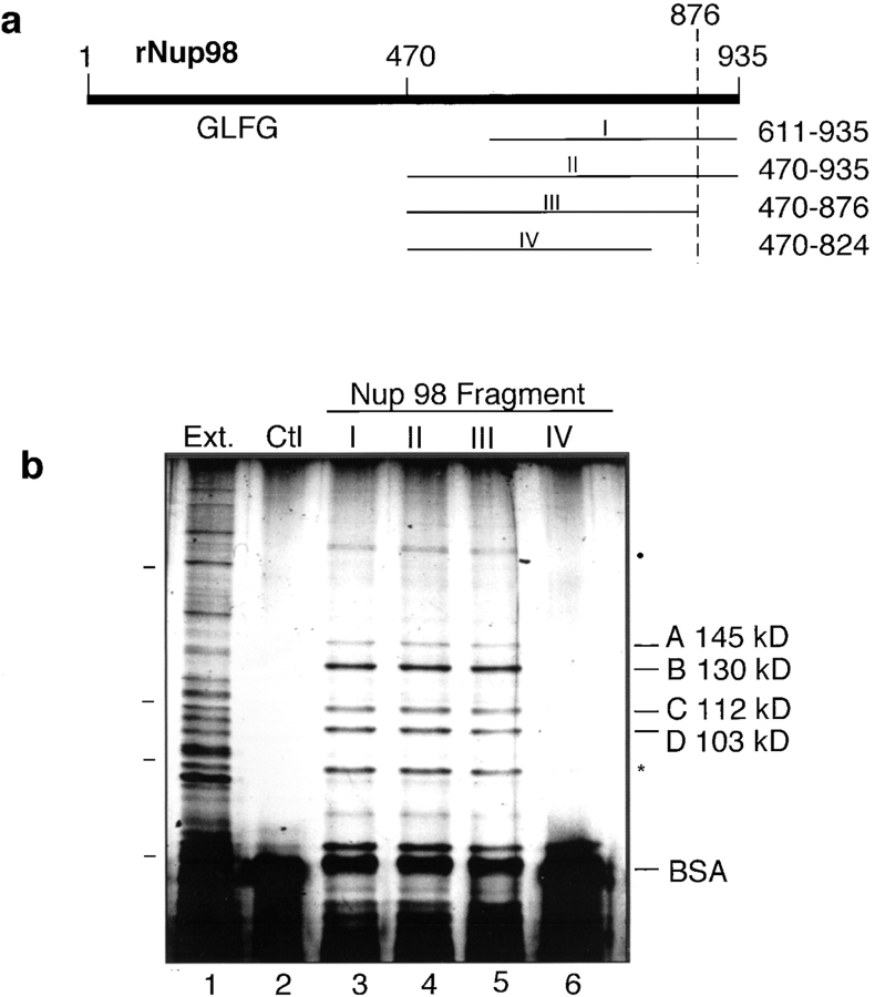
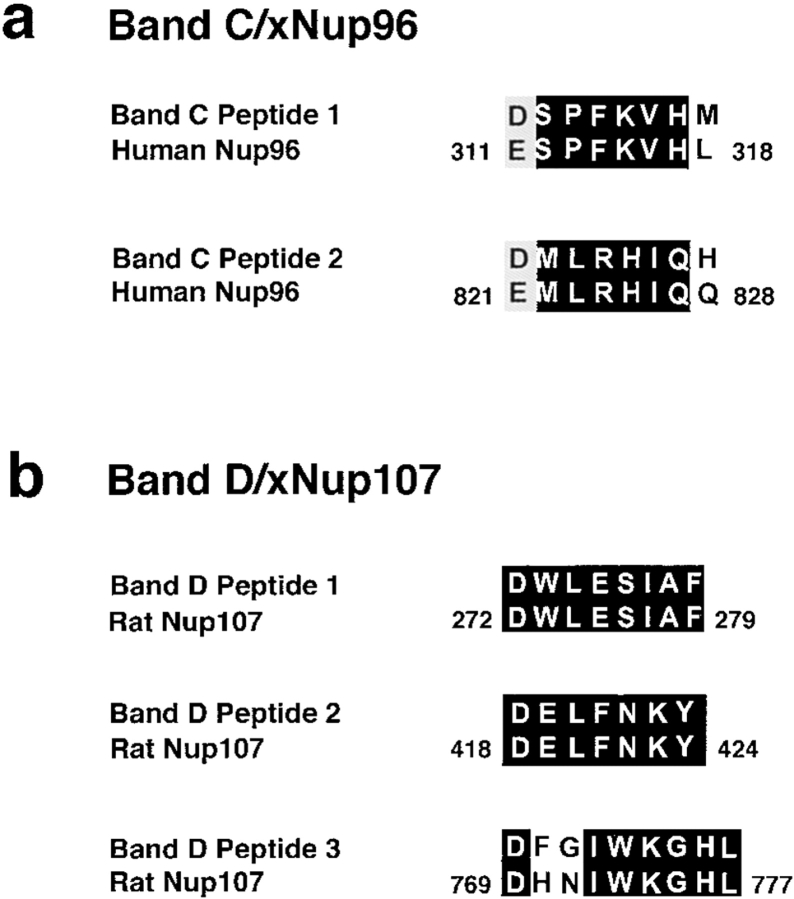
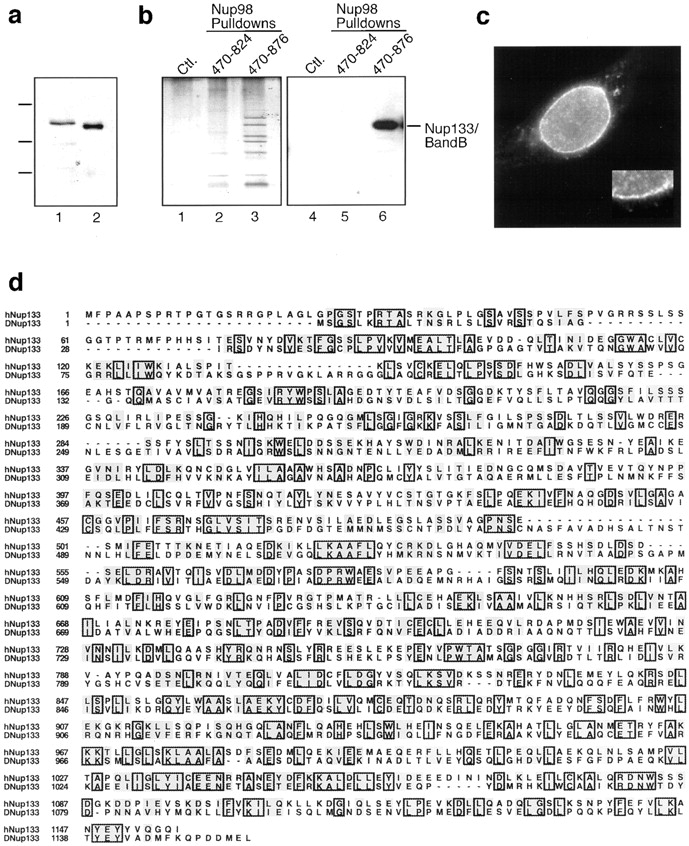
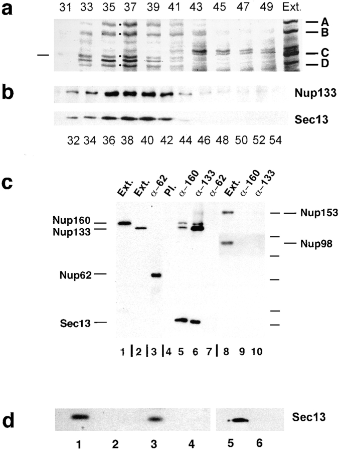
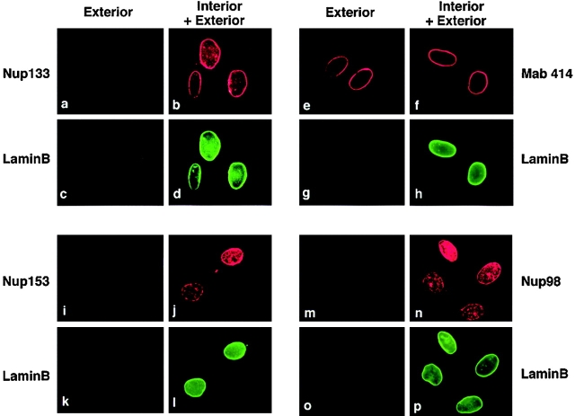
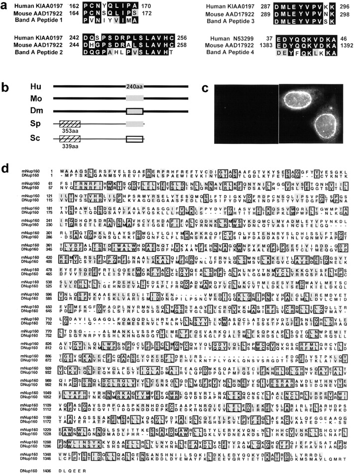
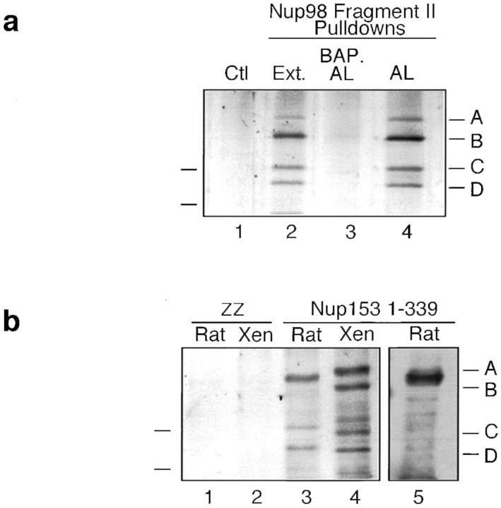
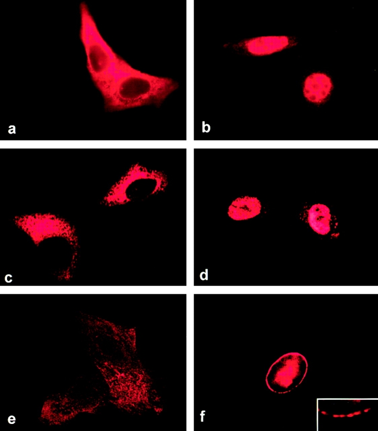
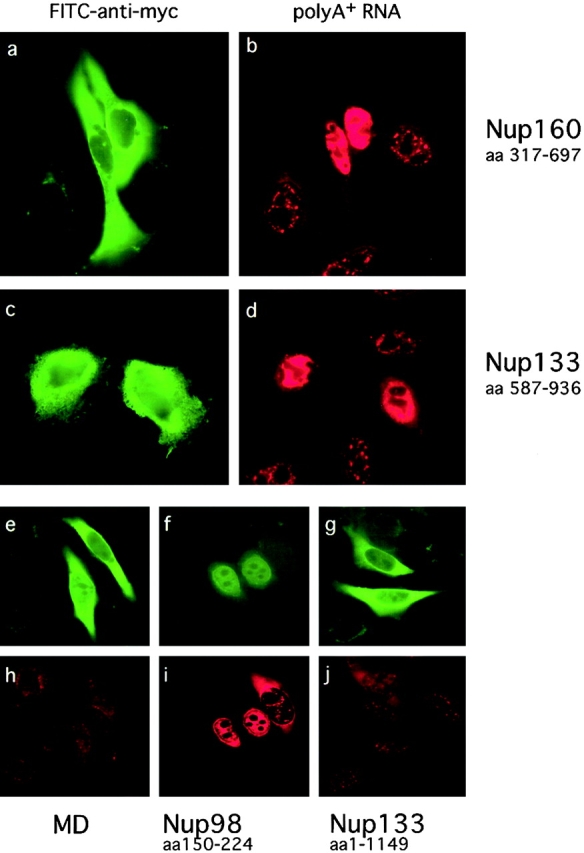
RNA undergoing nuclear export first encounters the basket of the nuclear pore. Two basket proteins, Nup98 and Nup153, are essential for mRNA export, but their molecular partners within the pore are largely unknown. Because the mechanism of RNA export will be in question as long as significant vertebrate pore proteins remain undiscovered, we set out to find their partners. Fragments of Nup98 and Nup153 were used for pulldown experiments from Xenopus egg extracts, which contain abundant disassembled nuclear pores. Strikingly, Nup98 and Nup153 each bound the same four large proteins. Purification and sequence analysis revealed that two are the known vertebrate nucleoporins, Nup96 and Nup107, whereas two mapped to ORFs of unknown function. The genes encoding the novel proteins were cloned, and antibodies were produced. Immunofluorescence reveals them to be new nucleoporins, designated Nup160 and Nup133, which are accessible on the basket side of the pore. Nucleoporins Nup160, Nup133, Nup107, and Nup96 exist as a complex in Xenopus egg extracts and in assembled pores, now termed the Nup160 complex. Sec13 is prominent in Nup98 and Nup153 pulldowns, and we find it to be a member of the Nup160 complex. We have mapped the sites that are required for binding the Nup160 subcomplex, and have found that in Nup98, the binding site is used to tether Nup98 to the nucleus; in Nup153, the binding site targets Nup153 to the nuclear pore. With transfection and in vivo transport assays, we find that specific Nup160 and Nup133 fragments block poly[A]+ RNA export, but not protein import or export. These results demonstrate that two novel vertebrate nucleoporins, Nup160 and Nup133, not only interact with Nup98 and Nup153, but themselves play a role in mRNA export.


Figures

Figure 2.
Figure 2.
Nucleoporin Nup153 binds the same four proteins. (a) A map of Nup153 and the fragments used. (b) Nup153 fragments coupled to beads were used in pulldowns. Nup153 aa 1–339 bound four proteins similar in size to A–D, termed A'–D' (lane 4). A zz tag control fragment and two Nup153 fragments, xNup153-N' and human Nup153 aa 1–245, did not (lanes 1–3). Size markers are 205, 116, 97, and 66 kd, respectively. (c) Nup98 fragment II and Nup153 aa 1–339 bind proteins identical in size (upper panel). Each also bound a protein of ∼97 kd (*; lanes 3 and 5) that was largely removed by the addition of RanQ69L (lanes 4 and 6). A–D and A'–D' binding were not sensitive to RanQ69L (lanes 3–6). Control S. aureus protein A-Sepharose beads did not bind A–D (lanes 1 and 2). RanQ69L was functional (lower panel), as it dissociated transportin from a Nup153 fragment (Shah and Forbes, 1998). The lower panel is an immunoblot with anti- transportin antibody; compare lane 5 (no Ran) with lane 6 (+Ran).
Figure 1.
Figure 1.
Proteins A–D bind to the COOH terminus of Nup98. (a) Subcloned fragments I–IV of rat Nup98. The dotted line shows the cleavage site for Nup98 endoproteolytic activity (Fontoura et al., 1999; Rosenblum and Blobel, 1999). Gle2 binds to Nup98 aa 150–224, whereas GLFG repeats occupy much of the remainder of aa 1–470. (b) Nup98 fragments I–IV coupled to beads were mixed with Xenopus egg cytosol in pulldown reactions. The * indicates a ∼97-kd Ran-sensitive protein. The filled circle indicates a band variably seen. Proteins A–D bound to fragments I–III (lanes 3–5), but not to fragment IV (lane 6) or S. aureus protein A beads (Ctl, lane 2). Xenopus egg extract (Ext; 0.02 μl) is shown for comparison (lane 1). Size markers are 205, 116, 97, and 66 kd, respectively (left hatchmarks).
Figure 3.
Figure 3.
Proteins C and D are the known vertebrate nucleoporins Nup96 and Nup107. (a) Two of the peptides obtained from Band C are shown. (b) Three of the peptides obtained from Band B and D are shown and match with near identity to rat nucleoporin Nup107. Identity is boxed and homology is indicated in gray, as defined by Kyte-Doolittle algorithms.
Figure 4.
Figure 4.
A novel vertebrate nucleoporin, Nup133. (a) Antibody to aa 777–1105 of the human protein AK001676, homologous to Xenopus protein B, recognized a single ∼130-kd protein in HeLa cell extracts (lane 1) and in rat liver nuclei (lane 2). Markers are 205, 116, and 97 kd. (b) Pulldowns with Nup98 fragments were split and electrophoresed on two gels. One was silver stained (lanes 1–3) and one immunoblotted with anti-Nup133 antibody (lanes 4–6). An antibody- reactive band of ∼130 kd was observed only in the Nup98 aa 470–876 pulldown (lane 6) and ran coincident with silver-stained protein B (lane 3). Ctl represents a pulldown with S. aureus protein A beads. (c) IF on HeLa cells using affinity-purified anti-Nup133 antibody; the inset shows a portion of the same nucleus magnified to reveal the punctate nuclear rim stain. (d) The sequence of the novel human nucleoporin Nup133 (AK001676) is compared with a highly related Drosophila homologue (AAF56042). Identity is boxed and homology is indicated in gray, as defined by Kyte-Doolittle algorithms.
Figure 7.
Figure 7.
The Nup160 nuclear pore subcomplex. (a) After gel filtration of Xenopus egg cytosol, odd fractions were subjected to Nup153 aa 1–339 pulldown, SDS-PAGE, and silver staining. A–D fractionated together and peaked at fraction 37 (dots). The left hatchmark indicates a 116-kd mw marker protein. When size controls were monitored for migration on the gel filtration column (unpublished data), Nup214 was seen to fractionate in a complex peaking at ∼1,000 kd (peak fraction = 34) and Nup98 fractionated in a complex peaking at ∼450 kd (peak fraction = 44), as expected (Macaulay et al., 1995). The dark band between C and D was not seen in pulldowns of unfractionated extract (Figs. 1 and 2) and is likely a breakdown product. (b) The even fractions (of total egg extract) were probed with anti-Nup133 and anti-sec13 antibodies. Nup133 peaks coincident with the peak of the silver-stained bands A–D indicated by dots in panel (a). The majority of soluble sec13 in Xenopus extracts also peaks in this region. (c) Immunoprecipitation from Xenopus cytosol using anti-xNup160 (lanes 5 and 9), anti-xNup133 (lanes 6 and 10), anti-xNup62 (lanes 3 and 7), and a mix of the preimmune sera for xNup160 and xNup133 (lane 4). The top portion of the blot corresponding to lanes 4–7 was probed with both anti-xNup160 and anti-xNup133 antibodies and the bottom with anti-sec13 antibody. The blot of lanes 8–10 was probed with both anti-rNup98 and anti-rNup153. Xenopus cytosol (0.2 μl) is shown in lanes 1, 2, and 8. Lane 1 was probed with anti-xNup160, lane 2 with anti-xNup133, and lane 3 with anti-xNup62. The molecular markers (left) were 200, 120, 90, 68, 53, 36, and 32 kd. Nup153 runs aberrantly high at ∼180 kd (Sukegawa and Blobel, 1993) (d) To ask whether sec13p is in Nup98 pulldowns, Nup98 aa 470–876 pulldowns were done from solubilized AL (lane 1), solubilized mock AL made in the presence of the pore assembly inhibitor BAPTA (lane 2), and egg extract (lane 3). A pulldown from egg extract using Nup98 aa 470–824 beads was done in lane 4. Pulldowns from Xenopus extract were also done with Nup153 aa 1–339 (lane 5) and Nup153 aa 1–245 (lane 6). All pulldowns were probed with anti–human sec13p antibody.
Figure 5.
Figure 5.
Antibody to Nup133 localizes to the nuclear side of the pore. HeLa cells were permeabilized with digitonin to allow antibodies access only to the exterior face of the nuclear envelope (Exterior). The monoclonal antibody mAb414 detects the presence of Nup214 and Nup358 on the cytoplasmic face of the nuclear pores (e). Nup133, Nup153, or Nup98 were not detected on this side of the pores (a, i, and m, respectively). As a control for nuclear envelope integrity, the cells were co-stained with the anti–lamin B antibody, LS-1 (c, g, k, and o). To probe the nuclear interior, Triton X-100 (Interior + Exterior) or longer digitonin (unpublished data) permeabilization was used. Anti–lamin B stain confirms nuclear envelope penetration of the antibody (d, h, l, and p) after TX-100. Nup153 and Nup98 are detected on the intranuclear face of pores, as expected (j and n). Nup133 is detected on the interior (b), but not the exterior (a) of the nuclear envelope.
Figure 6.
Figure 6.
A new vertebrate nucleoporin, Nup160. (a) Peptides from Xenopus band A match sequences of a 160- kd unknown mouse protein (AAD17922), a human clone (KIAA0197), and an overlapping human EST (N53299). (b) A schematic comparison of the human (Hu), mouse (Mo), and Drosophila (Dm) Nup160 proteins and distantly related S. cerevisiae ScNUP120 and putative S. pombe Nup120 proteins. Thick lines indicate relatedness, hatched boxes show relatedness between the two yeast proteins, gray boxes indicate a region of moderate homology between S. pombe and metazoans, and the black outlined box indicates a region of homology between ScNup120 and Drosophila Nup160. The hatched box and thin lines of the yeast proteins show little relatedness to the vertebrate proteins. (c) Antibody raised to the putative human Nup160 protein gives a punctate nuclear rim stain on TX-100 permeabilized HeLa cells (right panel), but not on digitonin-permeabilized cells (left panel), indicating localization of Nup160 on the basket face of the pore. (d) A comparison of mouse Nup160 and the highly related Drosophila homologue, aligned using Clustal W. Identities are boxed, homologies are in gray.
Figure 8.
Figure 8.
The Nup160 complex is in assembled pores. (a) AL assembly reactions were done in the presence or absence of BAPTA. AL were isolated and partially solubilized with 0.5 M NaCl which does not destabilize the A–D complex. Solubilized AL was clarified by centrifugation and used for pulldowns with Nup98 fragment II beads. Proteins A–D were pulled down from egg extract (lane 2, Ext) and from AL (lane 4) by Nup98 beads, but not by S. aureus protein A beads (lane 1, Ctl) or from AL reactions in which pore assembly had been inhibited by BAPTA (lane 3). Mw markers are 116 and 97 kd, respectively. (b) Rat liver nuclei were partially disassembled with 2% Triton. The clarified supernatant was incubated with Nup153 aa 1–339 beads. Three rat proteins in the relevant size range were observed by silver staining to be pulled down (lane 3), two similar to Xenopus C and D (lane 4), as well as a thick band intermediate between Xenopus A and B. None bound to zz-tag control beads (lanes 1 and 2). A portion of the Nup153 aa 1–339 pulldown from solubilized rat liver nuclei was probed with anti–human Nup133 antibody and gave a single immunoreactive band (lane 5) coincident with the thick band observed by silver stain (lane 3), indicating that this is rat Nup133.
Figure 9.
Figure 9.
Nup98 and Nup153 tether in the nucleus and to the pore, respectively, using their Nup160 complex binding domains. After transfection, myc-tagged Nup98 aa 470–824 is cytoplasmic (a and c), whereas Nup98 aa 470–876 is nuclear (b), like endogenous Nup98 (Powers et al., 1995). Nup98 aa 470–824 has access to the nucleus since it localizes there upon LMB addition (LMB; 100 nM, l hr) (d). Fragment 1C3 from the COOH terminus of Xenopus Nup153 (Shah et al., 1998) localized throughout the cell with no nuclear rim (e). Human Nup153 aa 1–339, which binds the Nup160 complex, localized to nuclear pores (f), as in Enarson et al. (1998).
Figure 10.
Figure 10.
Dominant negative fragments of Nup133 and Nup160 cause nuclear accumulation of poly[A] + RNA. HeLa cells were transfected with pCS2MT vectors containing: human Nup160 aa 317–697 (a and b) and 133 aa 587–936 (c and d), malate dehydrogenase (negative control; e and h), rat Nup98 aa 150–224 (positive inhibitory control; f and i), or human Nup133 aa 1–1149 (g and j) in myc-tagged form. Successful transfection was assayed by IF with FITC–anti-myc antibody (a, c, and e–g), whereas the localization of poly[A]+ RNA was assessed by hybridization of the coverslips with Texas red oligo[dT]50 (b, d, and h–j). Both human Nup160 aa 317–697 (b) and Nup133 aa 587–936 (d) caused a striking accumulation of poly[A]+ RNA in the nucleus, identical to that caused by Nup98 aa 150–224 (i).

Similar articles

Cited by

References

    1. Aitchison, J.D., G. Blobel, and M.P. Rout. 1995. Nup120p: a yeast nucleoporin required for NPC distribution and mRNA transport. J. Cell Biol. 131:1659–1675. - PMC - PubMed
    1. Allen, T.D., J.M. Cronshaw, S. Bagley, E. Kiseleva, and M.W. Goldberg. 2000. The nuclear pore complex: mediator of translocation between nucleus and cytoplasm. J. Cell Sci. 113:1651–1659. - PubMed
    1. Altschul, S.F., T.L. Madden, A.A. Schaffer, J. Zhang, Z. Zhang, W. Miller, and D.J. Lipman. 1997. Gapped BLAST and PSI-BLAST: a new generation of protein database search programs. Nucleic Acids Res. 25:3389–3402. - PMC - PubMed
    1. Bachi, A., I.C. Braun, J.P. Rodrigues, N. Pante, K. Ribbeck, C. von Kobbe, U. Kutay, M. Wilm, D. Gorlich, M. Carmo-Fonseca, and E. Izaurralde. 2000. The C-terminal domain of TAP interacts with the nuclear pore complex and promotes export of specific CTE-bearing RNA substrates. Rna. 6:136–158. - PMC - PubMed
    1. Bailer, S.M., S. Siniossoglou, A. Podtelejnikov, A. Hellwig, M. Mann, and E. Hurt. 1998. Nup116p and Nup100p are interchangeable through a conserved motif which constitutes a docking site for the mRNA transport factor Gle2p. Embo J. 17:1107–1119. - PMC - PubMed

Publication types

MeSH terms