. 2001 Oct 29;155(3):381-92.
doi: 10.1083/jcb.200105139. Epub 2001 Oct 29.

MyoD-positive myoblasts are present in mature fetal organs lacking skeletal muscle

Affiliations

MyoD-positive myoblasts are present in mature fetal organs lacking skeletal muscle

J Gerhart et al. J Cell Biol. .

Abstract

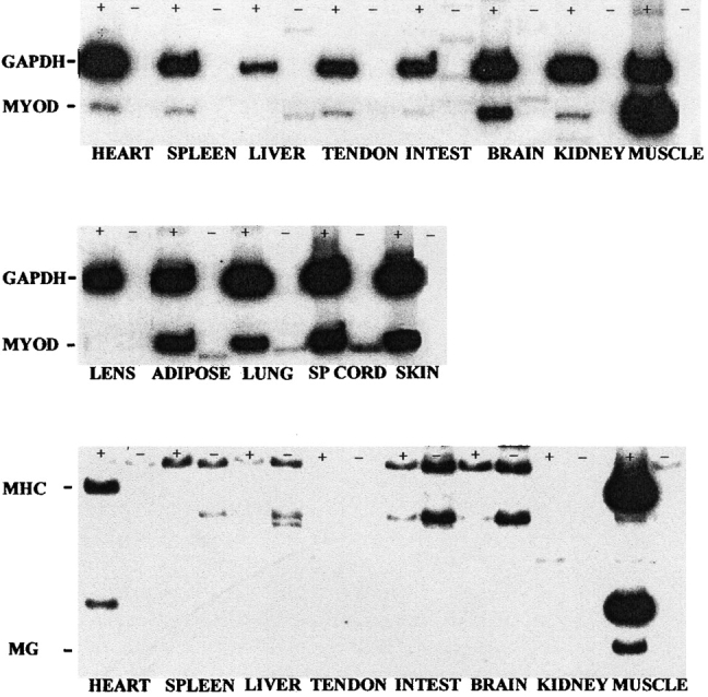
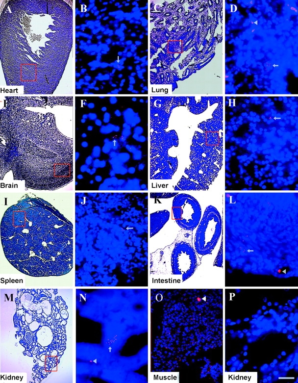
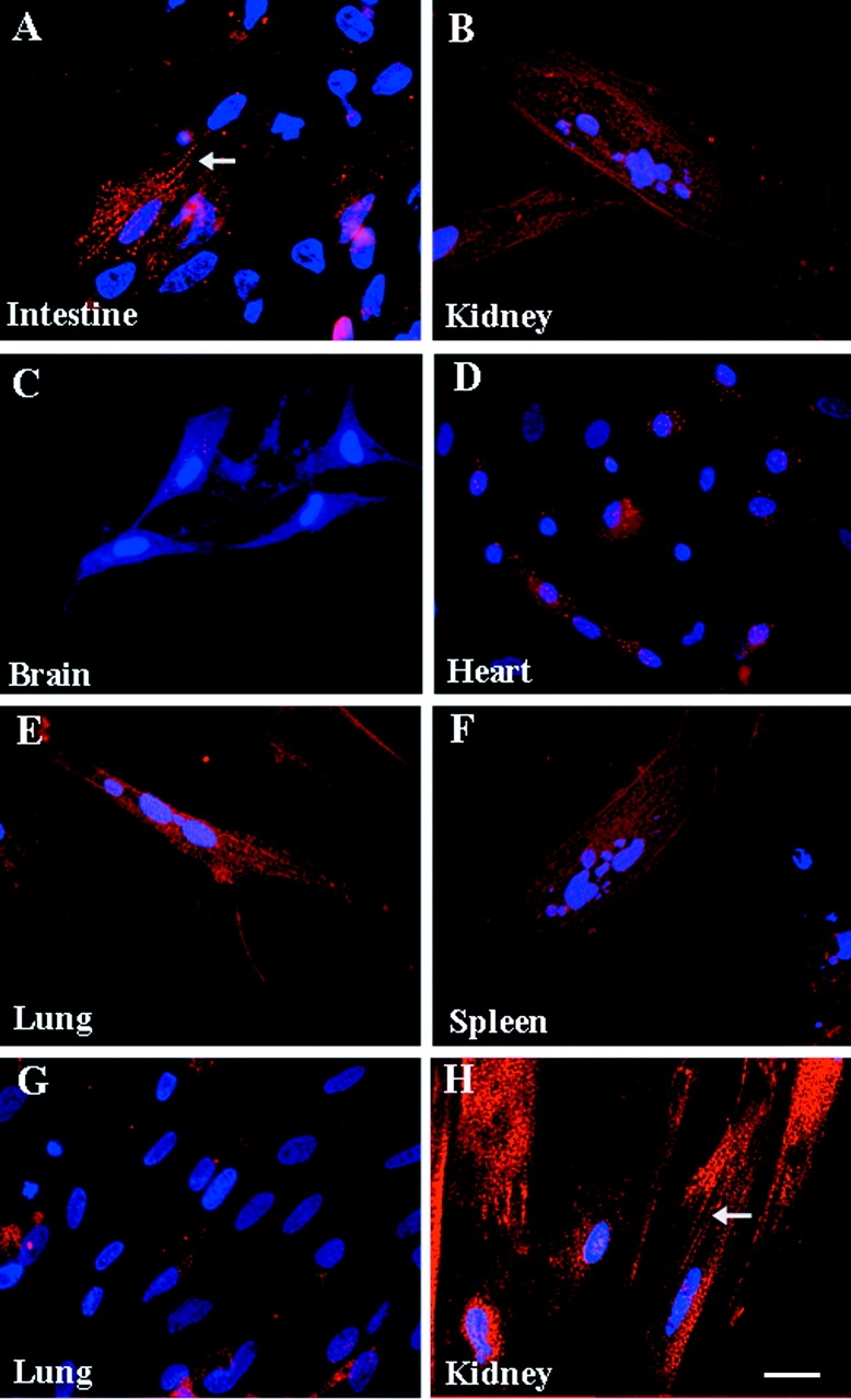
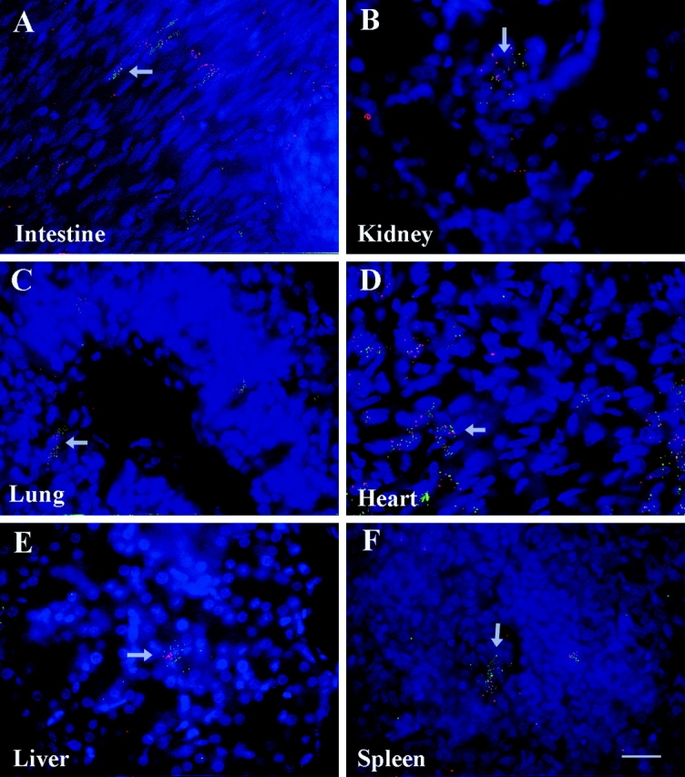
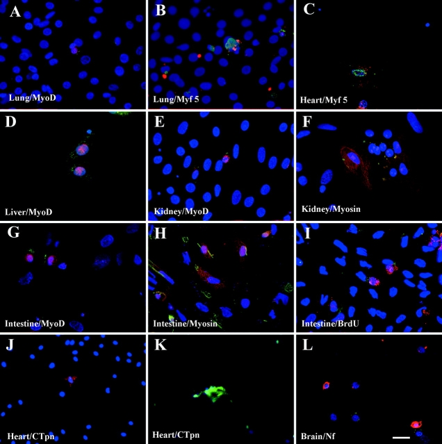
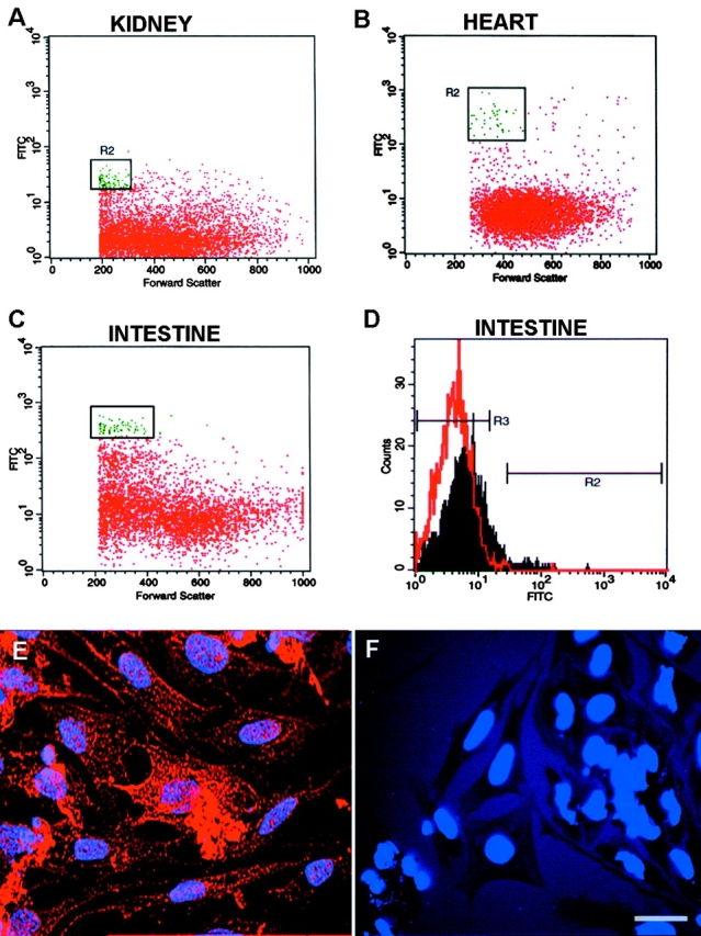
The epiblast of the chick embryo gives rise to the ectoderm, mesoderm, and endoderm during gastrulation. Previous studies revealed that MyoD-positive cells were present throughout the epiblast, suggesting that skeletal muscle precursors would become incorporated into all three germ layers. The focus of the present study was to examine a variety of organs from the chicken fetus for the presence of myogenic cells. RT-PCR and in situ hybridizations demonstrated that MyoD-positive cells were present in the brain, lung, intestine, kidney, spleen, heart, and liver. When these organs were dissociated and placed in culture, a subpopulation of cells differentiated into skeletal muscle. The G8 antibody was used to label those cells that expressed MyoD in vivo and to follow their fate in vitro. Most, if not all, of the muscle that formed in culture arose from cells that expressed MyoD and G8 in vivo. Practically all of the G8-positive cells from the intestine differentiated after purification by FACS. This population of ectopically located cells appears to be distinct from multipotential stem cells and myofibroblasts. They closely resemble quiescent, stably programmed skeletal myoblasts with the capacity to differentiate when placed in a permissive environment.


Figures

Figure 1.
Figure 1.
RT-PCR analysis of MyoD expression in fetal organs. mRNA from fetal organs was incubated with primers for MyoD, embryonic fast myosin (MHC), myogenin (MG), or GAPDH in the presence (+) or absence (−) of reverse transcriptase. MyoD (263 bp) was detected in all samples except the liver and lens. Myosin (616 bp) was expressed in pectoralis muscle and the heart. Myogenin (278 bp) was only detected in pectoralis muscle.
Figure 2.
Figure 2.
Localization of MyoD mRNA in fetal organs. In situ hybridizations were performed on sections of fetal organs. Low magnification images of the sections are shown to the left of each series. Fluorescence photomicrographs are the merged images of dendrimers in red and bis-benzamide labeled nuclei in blue. A few MyoD-positive cells were found in sections through each organ (arrows). Erythrocytes were autofluorescent (arrowheads). Pectoralis muscle was abundantly labeled with MyoD dendrimers (O). Dendrimers to myosin (P) did not label these sections. Bar, 135 μm in the fluorescence photomicrographs; 9 μm in the bright field images.
Figure 3.
Figure 3.
Myogenesis in cultures of cells prepared from fetal organs. Fetal organs were dissociated and the cells plated at high density. Cells were stained with the skeletal muscle specific 12101 (A–C, E and F) or troponin T (D) antibody, and an antibody to α-actinin (G and H). Striations can be seen in A and H (arrows). Multinucleated myotubes are shown in B and D–F. Bar, 9 μm.
Figure 4.
Figure 4.
Colocalization of the G8 antigen with MyoD mRNA in vivo. Sections were labeled with the G8 antibody and a secondary antibody conjugated with Alexa 488 (green). In situ hybridization was then performed with MyoD dendrimers (red). G8 colocalized with MyoD in most cells.
Figure 5.
Figure 5.
Immunofluorescence localization of cell type–specific proteins and BrdU in G8-positive cells in vitro. Cells from fetal organs were prelabeled with the G8 antibody, cultured for 48 h, and then stained with a secondary antibody to G8 and antibodies to MyoD, Myf5, or sarcomeric myosin. Cells from the lung, liver, kidney, and intestine that were labeled with G8 (green) had MyoD and myosin (red). Double labeling for G8 (red) and Myf5 (green) was seen in lung and heart cells. G8-positive intestine cells (green) incorporated BrdU (red) indicating replication. In a second type of experiment, cells from the heart and brain were fixed directly after removal from the fetus, centrifuged onto slides, and stained with G8 (red in J; green in L) and antibodies to cardiac troponin I (green) and neurofilament protein (red), respectively. Cells labeled with G8 did not have detectable levels of cardiac troponin (J) or neurofilament protein (L). Cells with cardiac troponin (K) and neurofilament protein (L) were not labeled with G8. Bar, 9 μm.
Figure 6.
Figure 6.
Flow cytometry and FACS ® of fetal cells. Fetal heart, kidney, and intestine cells were labeled with the G8 antibody and fluorescein-conjugated secondary antibody. Profiles of fluorescence intensity versus forward light scatter (cell size) were compared in cells labeled with G8 (green) and those labeled with the secondary antibody alone (red) (A–C). G8-positive (R2 population) and negative intestine cells (R3 population) were sorted (D), placed in culture for 2 d, and then stained with the 12101 antibody. Most of the G8-positive cells differentiated into skeletal muscle (E), whereas very few myosin positive cells were observed in the G8-negative cultures (F). Bar, 9 μm.

Similar articles

Cited by

References

    1. Bader, D., T. Masakki, and D.A. Fischman. 1982. Immunochemical analysis of myosin heavy chain during avian myogenesis in vivo and in vitro. J. Cell Biol. 95:763–770. - PMC - PubMed
    1. Barrofio, A., M.-L. Bochaton-Piallet, G. Gabbiani, and C.R. Bader. 1995. Heterogeneity in the progeny of single human muscle satellite cells. Differentiation. 59:259–268. - PubMed
    1. Beauchamp, J.R., J.E. Morgan, C.N. Pagel, and T. Partridge. 1999. Dynamics of myoblast transplantation reveal a discrete inority of precursors with stem cell–-like properties as the myogenic source. J. Cell Biol. 144:1113–1121. - PMC - PubMed
    1. Beauchamp, J.R., L. Heslop, D.S.W. Yu, S. Tajbakhsh, R.G. Kelly, A. Wernig, M.E. Buckingham, T.A. Partridge, and P.S. Zammit. 2000. Expression of CD34 and Myf5 defines the majority of quiescent adult skeletal muscle satellite cells. J. Cell Biol. 151:1221–1233. - PMC - PubMed
    1. Bellairs, R. 1986. The primitive streak. Anat. Embryol. 174:1–14. - PubMed

Publication types

MeSH terms