. 2001 Oct 29;155(3):405-14.
doi: 10.1083/jcb.200106141. Epub 2001 Oct 29.

Intraflagellar transport balances continuous turnover of outer doublet microtubules: implications for flagellar length control

Affiliations

Intraflagellar transport balances continuous turnover of outer doublet microtubules: implications for flagellar length control

W F Marshall et al. J Cell Biol. .

Abstract

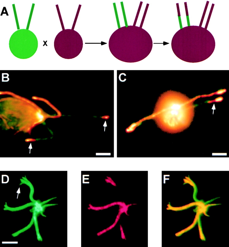
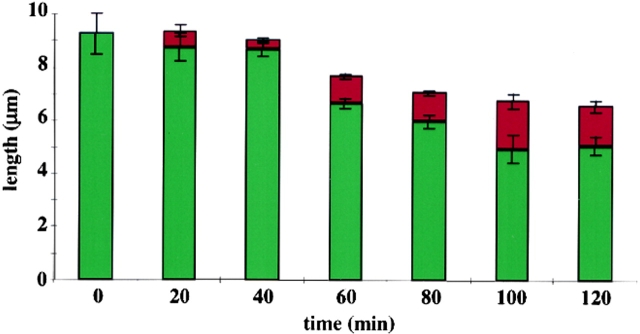
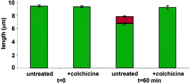
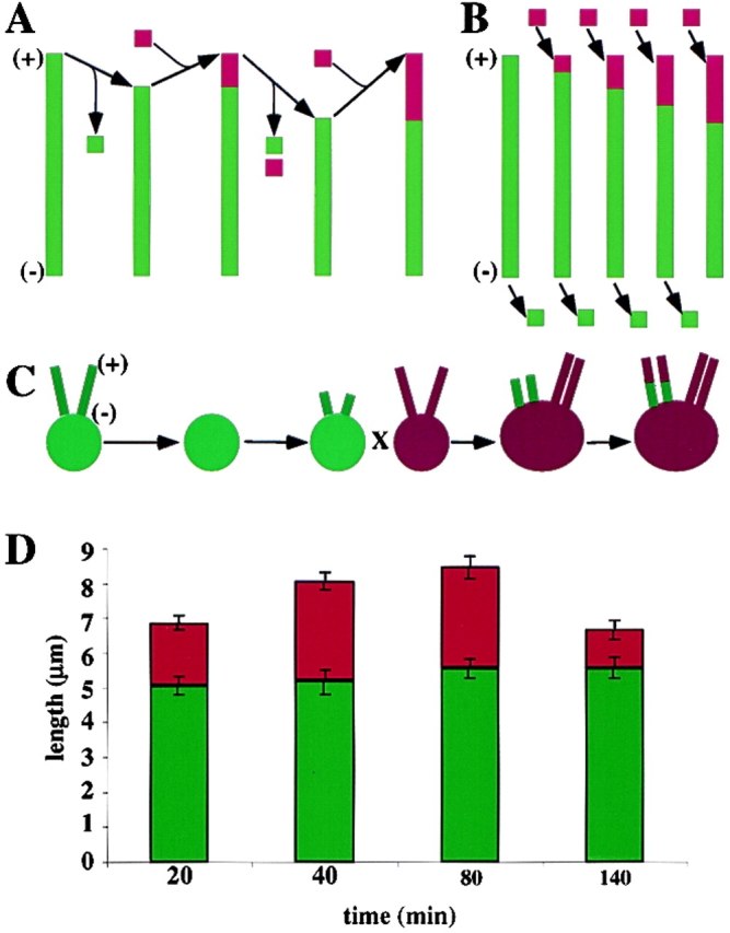
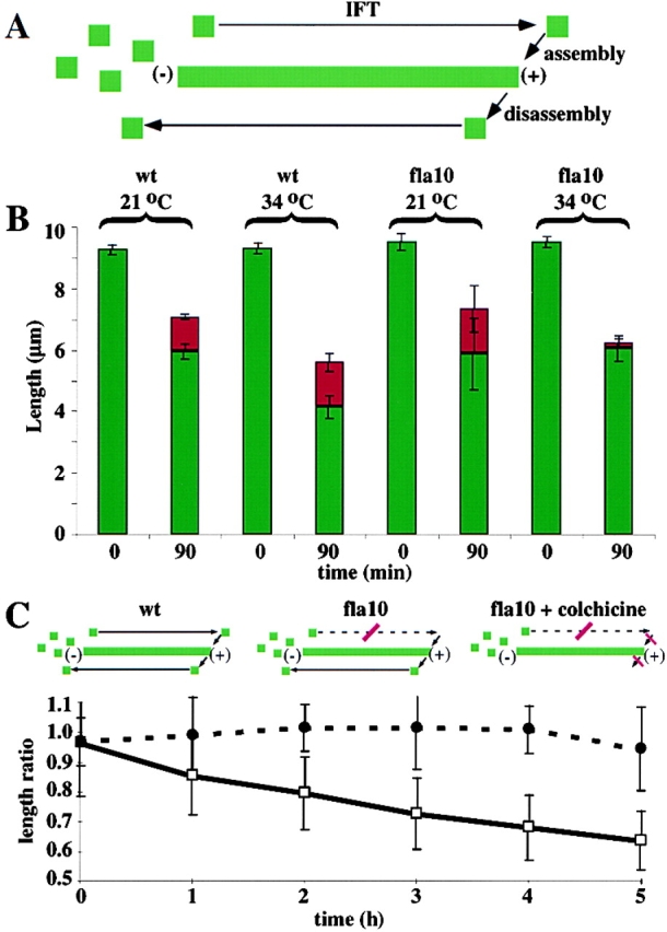
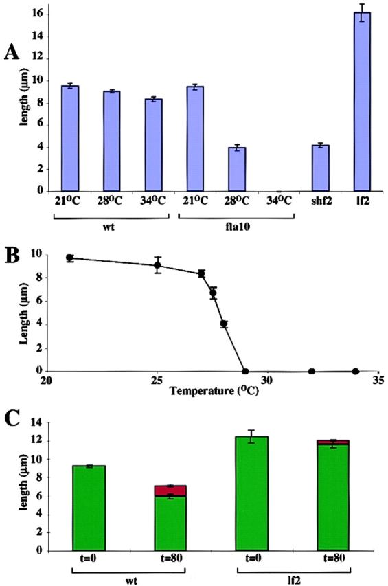
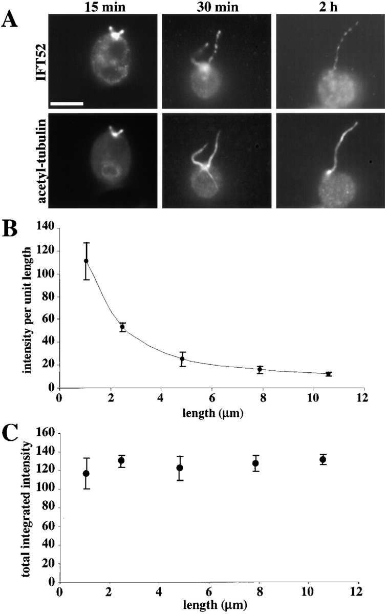
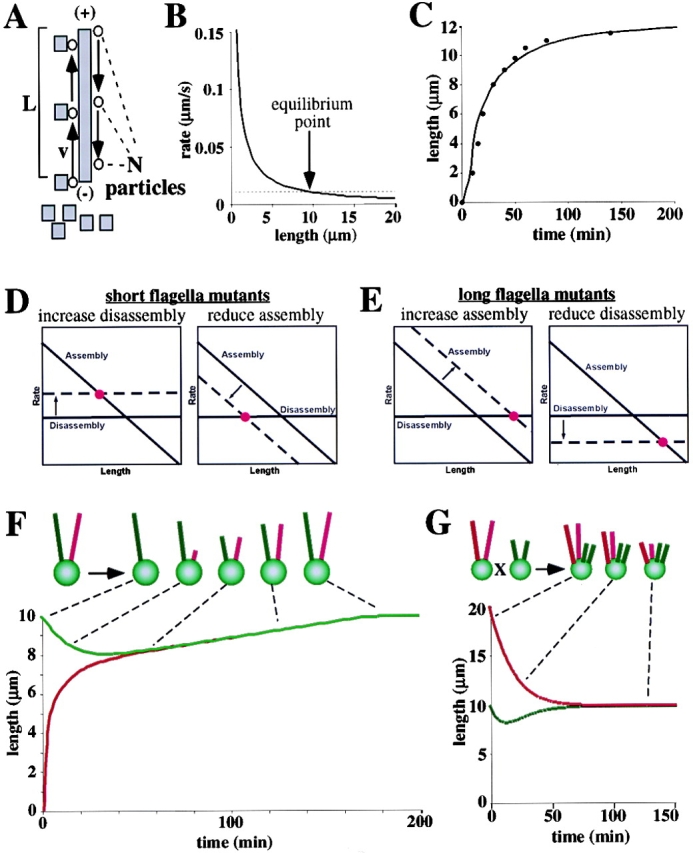
A central question in cell biology is how cells determine the size of their organelles. Flagellar length control is a convenient system for studying organelle size regulation. Mechanistic models proposed for flagellar length regulation have been constrained by the assumption that flagella are static structures once they are assembled. However, recent work has shown that flagella are dynamic and are constantly turning over. We have determined that this turnover occurs at the flagellar tips, and that the assembly portion of the turnover is mediated by intraflagellar transport (IFT). Blocking IFT inhibits the incorporation of tubulin at the flagellar tips and causes the flagella to resorb. These results lead to a simple steady-state model for flagellar length regulation by which a balance of assembly and disassembly can effectively regulate flagellar length.


Figures

Figure 1.
Figure 1.
Visualizing flagellar microtubule dynamics. (A) Experimental strategy. Cells expressing HA-tubulin are mated with nonexpressing cells to create a quadriflagellated dikaryon in which two flagella contain HA-tubulin and two do not. Cells are fixed at successive timepoints after mating, and stained with antibodies recognizing HA-tubulin, to detect incorporation of tagged tubulin into the previously unlabeled flagella. (B and C) Typical dikaryon images. Cell fixed 90 min after mating. (Green) FITC immunofluorescence with antibodies to Fla10 kinesin, used as a marker for flagella and basal bodies. (Red) Texas red immunofluorescence with antibodies recognizing the HA epitope–tagged tubulin. Yellow results from overlap of HA-tubulin staining with Fla10 kinesin staining, indicating incorporation of tagged tubulin into flagella. Two flagella (originally belonging to the tagged tubulin expressing cell) are fully labeled and two are partially labeled (arrows), indicating incorporation of tagged tubulin following mating. (D–F) Flagella splayed open with detergent plus ATP following mating of HA-tagged tubulin expressing and nonexpressing cells. (D) Splayed flagella detected by FITC immunofluorescence using antibodies against tubulin. Central pair microtubules (arrow) have extruded and are clearly separated from the main mass of outer doublets. (E) Texas red immunofluorescence using antibodies to HA epitope-tagged tubulin. Tagged tubulin clearly has incorporated into the distal end of the outer doublets, and possible also the splayed-out central pair microtubules. (F) Overlay image showing that majority of incorporation is in the outer doublet microtubules. Bars: (B and C) 2 μm; (D–F) 3 μm.
Figure 2.
Figure 2.
Measuring turnover based on spatial distribution of incorporated HA-tagged tubulin versus time. Each bar represents average labeling of flagella at a given time point after cell fusion. Red segment gives average length of labeled portion, green segment gives average length of unlabeled portion. Over time, HA-tagged tubulin incorporates over an increasingly large distal portion of the flagellum. At the same time, flagella are not growing but are in fact shortening as a consequence of the mating developmental pathway. Therefore, incorporation does not simply reflect residual flagellar growth, but must entail turnover of tubulin already present in the flagellum. Average number of flagella measured per time point was 27. Error bars in all graphs represent standard error of the mean.
Figure 3.
Figure 3.
Colchicine blocks flagellar tubulin turnover in wild-type cells. Compared to untreated cells, in cells treated with 3 mg/ml colchicine, a concentration that totally blocks flagellar regeneration, no assembly of HA-tubulin was observed at the distal ends of the flagella, confirming that the assembly observed in untreated cells represents bona fide microtubule polymerization. Colchicine also completely blocked disassembly of the outer doublets, thereby shutting down turnover. Average number of measurement per graph entry is 28.
Figure 4.
Figure 4.
Mechanism of flagellar microtubule turnover: plus end turnover versus treadmilling. (A) Plus end only turnover model. Individual outer doublet microtubules alternate between cycles of growth and shortening, with all dynamics restricted to the plus end. Plus and minus ends of flagellar doublets are indicated by (+) and (−). (B) Treadmilling model. New subunits are assembled at the plus end of the outer doublets, while simultaneously, subunits are disassembled from the minus end. (C) Experimental strategy to distinguish plus end turnover from treadmilling. Cells with regenerated half-length flagella were mated to cells expressing HA-tubulin. Right after mating, the half-length flagella rapidly elongate to full length, incorporating HA-tubulin along their distal half. (D) Result of partial regeneration experiment. The unlabeled proximal segment did not shorten, either during the rapid regeneration phase or during the subsequent steady-state phase. This result is inconsistent with a treadmilling mechanism, but matches the predictions of the plus end turnover model. Average number of flagella measured per time point was 10.
Figure 5.
Figure 5.
Role of IFT in flagellar maintenance. (A) Model: IFT particles carry structural proteins, including tubulin, to the plus end of the flagellum where they are incorporated in order to compensate for subunits removed during the disassembly portion of the turnover process. Plus and minus ends of the flagellar outer doublet microtubules are indicated by (+) and (−). (B) fla10 mutants at the nonpermissive temperature block the assembly portion of turnover, as judged by the lack of tagged tubulin incorporation at the tip, but do not block disassembly, as judged by shortening of the unlabeled region of the flagella. This confirms that IFT is needed for ongoing incorporation of new tubulin at the distal end. Average number of measurements per graph entry was 19. (C) Colchicine is predicted to rescue the fla10 phenotype by blocking outer doublet disassembly. As shown in graph, colchicine blocks flagellar resorption in fla10 cells shifted to the nonpermissive temperature. (Dotted line) fla10 cells treated with 3 mg/ml colchicine. (Solid line) Untreated fla10 cells. Length ratio plots ratio of length at nonpermissive temperature to length at permissive temperature.
Figure 6.
Figure 6.
Relationship between IFT, microtubule turnover, and flagellar length. (A) Partial reduction of IFT causes shf phenotype. Bars depict length of flagella in wild-type and fla10 cells grown at different temperatures. Typical shf and lf mutants are shown for comparison. In contrast to wild-type cells that show only a slight change in length at elevated temperatures, or temperature-sensitive fla10 cells grown at the nonpermissive temperature in which flagella are completely resorbed, when fla10 cells are grown at an intermediate temperature to partially reduce IFT, their flagella reach half length, comparable to bona fide shf mutants. (B) Length versus temperature in fla10 mutant cells grown at a range of temperatures spanning completely permissive and completely nonpermissive, resulting in intermediate steady state length. Average number of measurements per data point is 25. (C) Reduction in microtubule turnover rate in lf mutant. Compared to wild-type cells measured over an equal time interval, lf mutant lf2-5 shows less incorporation of new tubulin (reflected by the shorter length of incorporated tubulin at the plus end, indicated in red), as well as less removal of unlabeled tubulin (reflected by a smaller reduction in the length of the unlabeled proximal segment, indicated in green). Average number of measurements per entry in graph was 23.
Figure 7.
Figure 7.
Total quantity of intraflagellar transport particles is independent of flagellar length. (A) IFT52 immunofluorescence staining during flagellar regeneration. Three cells are shown at three different time points during regeneration. As regeneration progresses and flagella elongate, the density of IFT52 decreases visibly. Bar, 5 μm. (B) Average IFT52 staining intensity, per unit length, plotted versus flagellar length. Quantification by digital imaging confirms the visual impression that IFT particle staining is more intense in short flagella. (C) Total IFT52 fluorescence intensity, a measure of IFT particle protein content, found by multiplying intensity per unit length by flagellar length. Graph indicates that the IFT particle content of a flagellum is independent of flagellar length.
Figure 8.
Figure 8.
Theoretical model for flagellar length control based on IFT and microtubule turnover. (A) IFT leads to length-dependent assembly rate. Equation gives assembly versus length assuming that transport is rate limiting. N number of IFT particles cycling per flagellum. α, Constant reflecting the transport capacity of a single IFT particle; v, average velocity of an IFT particle; L, flagellar length. Squares represent flagellar precursor protein, for example tubulin. Circles represent IFT particles. (B) Length-dependent assembly predicted by IFT-limited transport leads to a steady-state mechanism for length regulation. (Solid line) Length-dependent assembly rate. (Dotted line) Length-independent disassembly rate. The flagellar length is the equilibrium length (arrow) at which the two rates balance. (C) Predicted kinetics of flagellar regeneration based on equation 1. (•) Data points taken from regeneration measurements in Rosenbaum et al. (1969). (Solid line) Best fit predicted regeneration kinetics. (D) Two possible causes of shf mutants. Either increasing disassembly rate (left) or decreasing assembly rate (right) will lead to equilibration at a shorter length. Thus, reduction in IFT is predicted to cause an shf phenotype, which accounts for the results of Fig. 6 A. (Solid line) Wild-type rates; (dotted line) rate in mutant cells; (•) new equilibrium point in mutant. (E) Two possible causes of lf mutants. Either increasing assembly rate (left) or decreasing disassembly rate (right) will lead to equilibration at a longer length. The ability of reduced turnover to cause lf phenotype accounts for the results of Fig. 6 C. (F) Turnover model predicts results of experiment in which a single flagellum is severed. Cartoon illustrates observed behavior of such cells: as the severed flagellum regenerates, the intact flagellum shortens until both reach the same length. Graph shows result of computer simulation based on length control model. (Red) Length of severed flagellum; (green) length of the unsevered flagellum. Dashed lines connect corresponding time points of simulated response with stages of observed response. (G) Simulation of experiments in which recessive lf mutants are fused with wild-type cells. (Red) lf shorten to wild-type length once the recessive mutation is complemented by wild-type cytoplasm; (green) wild-type flagella fluctuate in length, but eventually regain their normal length.

References

    1. Asleson, C.M., and P.A. Lefebvre. 1998. Genetic analysis of flagellar length control in Chlamydomonas reinhardtii: a new long-flagella locus and extragenic suppressor mutations. Genetics. 148:693–702. - PMC - PubMed
    1. Barsel, S.E., D.E. Wexler, and P.A. Lefebvre. 1988. Genetic analysis of long-flagella mutants of Chlamydomonas reinhardtii. Genetics. 118:637–648. - PMC - PubMed
    1. Behnke, O., and A. Forer. 1967. Evidence for four classes of microtubules in individual cells. J. Cell Sci. 2:169–192. - PubMed
    1. Cole, D.G., D.R. Diener, A.L. Himelblau, P.L. Beech, J.C. Fuster, and J.L. Rosenbaum. 1998. Chlamydomonas kinesin-II–dependent intraflagellar transport (IFT): IFT particles contain proteins required for ciliary assembly in Caenorhabditis elegans sensory cilia. J. Cell Biol. 141:993–1008. - PMC - PubMed
    1. Coyne, B., and J.L. Rosenbaum. 1970. Flagellar elongation and shortening in Chlamydomonas. II. Reutilization of flagellar proteins. J. Cell Biol. 47:777–781. - PMC - PubMed

Publication types