Case Reports
. 2001 Nov;54(11):887-9.
doi: 10.1136/jcp.54.11.887.

Post-transplant malignant lymphoma with monoclonal immunoglobulin gene rearrangement and polyclonal Epstein-Barr virus episomes

Affiliations
Case Reports

Post-transplant malignant lymphoma with monoclonal immunoglobulin gene rearrangement and polyclonal Epstein-Barr virus episomes

Y Sadahira et al. J Clin Pathol. 2001 Nov.

Abstract

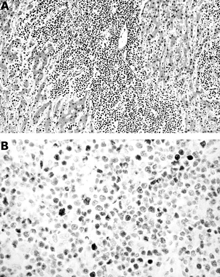
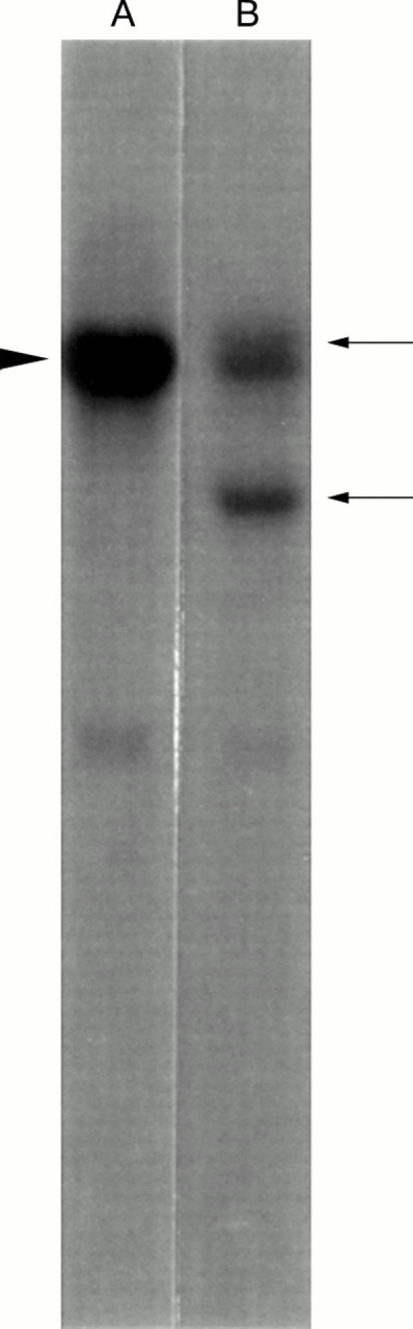
This report describes the case of an 8 year old boy who developed ileocecal B cell lymphoma after liver transplantation. The patient underwent orthotopic liver transplantation for biliary atresia and had been given immunosuppressive drugs--cyclosporin A and tacrolimus hydrate. Six years after the liver transplantation, the patient had a sudden onset of fever and abdominal pain. Necropsy revealed an ileocecal mass that was a B cell lymphoma. Epstein-Barr virus (EBV) encoded RNA 1 was demonstrated in lymphoma cells and hyperplastic follicular germinal centre cells in various tissues. Although monoclonal immunoglobulin gene rearrangement was detected in the liver, EBV episomes were of polyclonal origin and lytic forms of EBV were also demonstrated by Southern blotting. Immunohistochemically, lymphoma cells were positive for p53 but negative for latent membrane protein 1 and EBV nuclear antigen 2. These findings suggested that this B cell lymphoma might have occurred sporadically, regardless of EBV infection.


Figures

None
Figure 1 (A) Lymphoma cell infiltration of the liver. Haematoxylin and eosin staining; original magnification, x35. (B) Immunostaining for p53. The nuclei of lymphoma cells are positive. Original magnification, x140.
None
Figure 2 Southern blot analyses of liver with the JH probe after digestion with HindIII. Lane A, placental DNA showing a germ line band (arrowhead); lane B, DNA from the liver. Arrows show rearranged bands.
None
Figure 3 PCR amplification of the Fr2-JH region of the rearranged immunoglobulin gene in the ileocecal tumour (lane A) and the liver (lane B) using paraffin wax embedded sections. PCR primers were prepared using a DNA synthesiser, as described previously.8 The PCR products were separated on an agarose gel containing 0.3 mg/ml (0.003%) of ethidium bromide, and bands were visualised and photographed using ultraviolet transillumination. The predicted sizes of the amplified products were 240–280 bp (arrow). Lane C, positive control (a paraffin wax section of diffuse large cell lymphoma); lane D, negative control (an anaplastic large cell lymphoma cell line); lane E, molecular weight markers (a 100 bp ladder marker).
None
Figure 4 Epstein Barr virus (EBV) infection of the liver demonstrated using EBV encoded RNA 1 (EBER-1) in situ hybridisation. Nuclei of lymphoma cells were EBER-1 positive, whereas hepatic cells were negative. Original magnification, x140.
None
Figure 5 Southern blot analysis of the liver. For the presence of latent episomal or linear replication Epstein Barr virus (EBV) DNA, the 7.7 kb BamHI NJhet fused genomic terminal fragment was used as a probe. Lane A, molecular marker; lane B, negative control; lane C, positive control, containing DNA from an EBV infected polyclonal lymphoblastoid cell line (B95–8), which is permissive for lytic EBV infection. Both circular and linear forms of the virus genome are seen. Lane D, DNA from the liver, containing multiple high molecular bands consistent with episomal viral DNA and additional bands in the range of 2.5 to 3 kb consistent with linear viral DNA.

Publication types

MeSH terms