. 2001 Nov;234(5):607-12.
doi: 10.1097/00000658-200111000-00004.

Endoscopic suturing and knot tying: theory into practice

Affiliations

Endoscopic suturing and knot tying: theory into practice

D L Murphy. Ann Surg. 2001 Nov.

Abstract

Objective: To advance modern surgical techniques of endoscopic knot tying, encompassing a new appreciation of knot-tying theory and the application of second-generation, purpose-designed instruments.

Summary background data: During open surgery, surgeons automatically create the surgical half-hitch by using either instrument or hand/finger knot-tying methods (figure 4). Each of these methods, which are mirror images of each other, forms the same result, the half-hitch. Two opposing half-hitches are needed to form a square knot. There are many ways for new-generation instruments to create a secure square knot during endoscopic surgery. An overview of the current endoscopic knot-tying methods is presented.

Methods: The author presents a theoretical analysis of square knot-tying techniques as applied during instrument and hand/finger movements. The application of a mirror-image concept was considered in the analysis of these two contrasting methods.

Results: There are 12 ways to create a square knot, some of which have previously not been described or needed in open surgery. Some of these methods have particular application in endoscopic surgery.

Conclusions: A new understanding of knot-tying theory has been developed, with innovative methods being defined for tissue approximation during endoscopic surgery. These ergonomic, efficient, and contrasting methods of knot tying are described using second-generation endoscopic instruments. The new techniques have direct and broad application in many fields of minimally invasive surgery.


Figures
None
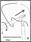
Figure 1. Instrument reverse loop.
None
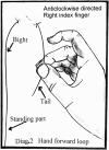
Figure 2. Hand forward loop.
None
Figure 3. New instrument.
None
Figure 4. Diagrammatic display of how the jaw and spur operate to form a half hitch. (A) Spur action alone. The role of the right instrument and appendage spur is shown capturing and controlling the bight to form forward-directed loops. (B) Combined jaw and spur action. (a) The jaws of the right instrument hold the suture tail. (b) With forward right instrument movement and counterclockwise rotation, the spur appendage of this instrument captures and directionally controls the bight section of the suture. (c) One full rotation of right. (d) One-and-a-half rotations, then through the loop, carrying the suture tail to complete a forward-directed loop of a half-hitch. Multiple instrument rotations are easily performed to create extra loops, as in a surgical half-hitch. First-generation endoscopic needle holders do not easily perform this task. The partner left instrument (not shown) in turn holds the standing part of the suture coaxially with the rotating right instrument.
None
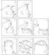
Figure 5. Open-surgery method. (a) Configuration of a C formation on the bight with the right. (b) Left clockwise rotation and advance to form the reverse-directed loop on the suture. (c) Left grasps the tail, rotated counterclockwise and withdrawn. (d) The first half-hitch. (e) Left transferred and holds the suture in a C-loop configuration. Right is released from bight, and the jaw is closed. Next counterclockwise rotation of right around the suture forms the second reverse loop on the bight. (f) Right grasps the tail. (g) Right rotates clockwise and withdraws the tail. (h) Completed square knot. Instrument separation tightens the knot.
None
Figure 6. Hand-tie method, performed with instruments. (a) Right holds tail; left holds bight. (b) Right counterclockwise rotation over the bight; the spur captures the suture to form a loop under the bight. Clockwise rotation of left can assist in this loop formation. (c) Further right counterclockwise rotation under bight and through the loop. A double-loop, surgical half-hitch can easily be formed by further rotation of right around the bight. Left transfers to grasp the tail. (d) Release suture tail from right, close right jaw, then clockwise rotation and withdrawal. (e) Right grasps bight; separation of right and left tightens the half hitch. (f) Counterclockwise rotation of right forms a loop on the bight; clockwise rotation of left over bight and advance through the loop. The spur appendage can assist with this loop formation. Next (not shown), right transfers to grasp tail. Repeat (d) and (e) with the opposite instruments to complete and tighten the square knot. Further half-hitch loops can be performed as needed.
None
Figure 7. Combination of instrument and hand-tie methods. Only one instrument interchange is needed. (a) Left holds bight in C formation. (b) Right orbital clockwise rotation under the bight forms a reverse loop, or (c) two or more loops. (d) Right grasps tail, followed by clockwise rotation and withdrawal of right. (e) First half-hitch formed; instrument separation tightens the hitch. Specific instrument movements, as in open surgery, can create suture-locking techniques if needed. (f) Right counterclockwise rotation over then forward and under the bight creates a forward loop. The spur can assist with this loop formation, as does clockwise rotation of left. (g) Left transferred to tail, release right, close jaw, and clockwise rotational withdrawal of right. (h) Right grasps bight; instrument separation tightens the square knot.
None
Figure 8. Instrument tie versus hand tie.
None
Figure 9. Instrument tie versus hand-tie method using instruments.

References

    1. Cuschieri A, Szabo Z. Tissue approximation in endoscopic surgery. ISIS Medical Medial, 1995.
    1. Ferzli GS. Laparoscopic Instrument. U.S. Patent 005147 373, 1992.
    1. Noda WA. Dual Function Suturing Device, Method. U.S. Patent 005211650A, 1993.
    1. Murphy DL. Endoscopic knot tying made easier. Aust NZ J Surg 1995; 65: 507–509. - PubMed
    1. Murphy DL. A new method of laparoscopic instrument knot tying. Surgical Technology International IV 1995; 4: 199–202. - PubMed